Magic Tooth Dental Center      +962 6 593 7076     +962 6 593 6367

Magic Tooth Dental CenterDental implant patient guide

Dental implants in Amman, Jordan, explained one step at a time

At Magic Tooth Dental Center in Abdoun, Amman, Jordan, replacing a missing tooth with a dental implant happens in stages. These eight guides explain what we use at each stage, from the 3D scan to your new crown, and what it means for you. Read them in order, or go straight to the part you want to know about.

3D imaging at Magic Tooth Dental Center

See the full picture before your implant

A dental implant has to sit in solid bone, clear of nerves and sinuses. So before we place one, we take a 3D X-ray of your jaw, called a CBCT scan, on our Castellini X-Radius Trio. Your dentist can then measure your bone and choose the implant’s size and position on screen first.

  • Painless
  • Over in under a minute
  • Done here in our clinic

What your dentist sees

A flat picture shows how tall the bone is, but not how thick.

  • Bone height. Can be seen.
  • Nerve canal. Seen from the side only.
  • Bone thickness. Hidden, so this view alone cannot show whether an implant will fit.
A slice through the lower jaw from a 3D scan A cross-section of the jawbone with an implant and a new tooth in the top of the bone. An arrow marks 14.5 millimetres of bone height above the nerve canal, a line marks a bone width of 6.8 millimetres, and the nerve canal appears as a dark circle lower down. A B C D

A 3D scan can be sliced, so the bone’s thickness and the nerve’s position come into view.

  • Bone width. Measured here as 6.8 mm.
  • Bone height. Measured here as 14.5 mm above the nerve.
  • Nerve canal. Carries the nerve to your lip and chin.
  • Implant and new tooth. Sized and angled to stay clear of the nerve.

Select a letter to highlight it in the picture.

Example images, for illustration.

What is a CBCT scan?

CBCT stands for cone beam computed tomography. The scanner’s arm moves around your head while sending a cone-shaped X-ray beam through your jaw. A computer then turns what it captures into a three-dimensional picture of your teeth, your jawbone, the bony canals that carry your nerves, and your sinuses.1

The scan is quick and painless, and it is done here in the clinic. Dentists use it to plan implants and for other situations where a flat X-ray cannot answer the question.1

An ordinary X-ray is like a flat map

You can see where the streets are, but not how tall the buildings stand. Everything is flattened into one picture.

A CBCT scan is like a globe you can turn

Your dentist can rotate it, zoom in, and look through your jaw one thin slice at a time, from any direction.

Why 3D matters for your implant

An implant is a small titanium post that replaces the root of a missing tooth. Where it goes, how long it is and the angle it sits at all depend on what lies beneath your gum.

  • It measures your bone

    An implant needs enough bone all the way around it. The scan shows the bone’s height, width and shape, so your dentist knows before surgery whether an implant will fit.23

  • It shows where the nerve runs

    In the lower jaw, the nerve that gives feeling to your lip and chin runs through a canal in the bone. Seeing that canal in 3D lets your dentist plan an implant that stays clear of it.23

  • It shows how close the sinus is

    Above your upper back teeth are air-filled spaces called sinuses. The scan shows how much bone lies beneath them.3

  • It lets your dentist rehearse on screen

    Your dentist can place a virtual implant on the scan and try different sizes, depths and angles, to suit both your bone and the new tooth that will sit on top.34

  • It shows if extra bone is needed

    If the bone is thin or shallow, the scan helps your dentist decide whether a bone graft or a sinus lift is needed before the implant, or alongside it.2

  • It means fewer surprises on the day

    Some things are easy to miss on a flat picture, such as an extra nerve opening or a narrow ridge of bone. It is far better to find them while planning than during surgery.3

The American Academy of Oral and Maxillofacial Radiology recommends 3D cross-sectional imaging for any potential implant site, and names CBCT as the method of choice.2

The American Dental Association’s 2026 recommendations also describe CBCT as the scan used to plan implant surgery.5

Our scanner: the Castellini X-Radius Trio

Castellini is an Italian maker of dental equipment, founded in 1935. Our X-Radius Trio takes ordinary panoramic X-rays and 3D CBCT scans on one machine. Here is what its features mean for you.6

It scans only the area that is needed

Choose a scan size to see what it covers.

One implant. The scan can be kept to the teeth and bone around the implant site.

Several implants in one jaw. The scan covers that whole jaw.

A plan for the whole mouth. One scan can take in both jaws.

The blue box is the area scanned and the red outline marks the implant site. A smaller area means less of you is exposed to X-rays.12

  • Exposure matched to the job

    The X-ray beam is pulsed and there are different scan modes, so your dentist can choose the setting that gives enough detail for your case and no more.

  • A quick check before the scan

    A head support and guide lights help line you up, and a preview image confirms the scan is aimed at the right area. That helps avoid a repeat.

  • Fine detail

    The 3D picture is built from tiny cubes, like the pixels in a photo. On this scanner they can be as small as 0.075 mm, less than a tenth of a millimetre.

  • Open and quick

    You stand in an open scanner, not inside a tunnel, and the scan itself usually takes less than a minute.7

  • Planning software

    The scan opens in Castellini’s iRYS software. Your dentist can place a virtual implant, see how far it sits from the nerve canal and set a safety distance. Scans are stored in DICOM, the standard format for medical images.4

How CBCT compares

No single scan is best for everything. Each one answers a different question. Choose one to compare it with CBCT.

What CBCT adds

  • It shows how thick the bone is. A flat X-ray cannot.23
  • It shows the nerve canal and the sinus in 3D. A flat X-ray shows them from one side only, so depth cannot be judged.2
  • It lets your dentist plan the implant’s position on screen.3

Where ordinary X-rays are better

  • They use less radiation.1
  • They are the right choice for check-ups and for the first look before an implant.25
  • They are usually enough for routine checks on an implant that is causing no trouble.2

What CBCT offers

  • It is designed for teeth and jaws, and gives the 3D bone detail an implant plan needs.3
  • It generally uses less radiation than a conventional CT scan.17
  • It is done in the dental clinic, while you stand.

Where a hospital CT is better

  • It shows soft tissues such as muscles and glands more clearly.7
  • Doctors use it for medical questions that go beyond teeth and jawbone.7

What our X-Radius Trio offers

  • A choice of scan sizes, so the scan can be kept to the area your dentist needs.6
  • Positioning aids and a preview check, to help get the scan right first time.6
  • A direct path from the scan to implant planning software.4

Worth knowing

  • Other modern CBCT scanners offer similar features.
  • Dose and image quality vary a great deal between machines, and with the scan size and settings chosen.38
  • What matters most is that the scan is tailored to you: the smallest area and the lowest dose that still give a clear enough picture.3

A scan supports good planning. It does not replace your examination, the treatment itself or the care you give your implant afterwards.9

Radiation and safety

A CBCT scan uses X-rays, so it is fair to ask about radiation. Here is the plain picture.

Where CBCT sits

  1. Ordinary dental X-raylowest dose
  2. Dental CBCTin between
  3. Hospital CTgenerally highest

This shows the order only. The steps are not to scale.17

  • Roughly two weeks to a month of everyday background radiation

    A review of measurements from many different CBCT machines found average adult doses of about 84 microsieverts for small scans and about 212 for large ones.8 Everyone receives around 2,400 microsieverts a year from natural sources such as the ground, the air and space.10 These are averages across machines, not a measurement of our scanner, and doses vary widely with the machine and the settings used.

  • A scan chosen for a reason

    Your dentist examines you first and looks at any earlier X-rays. A CBCT scan is taken only when the extra information will help your treatment, and then its benefit outweighs the small radiation risk.111

  • The smallest area that will do

    The scan is kept as small as it can be while still including what your dentist needs to see.12

  • Settings matched to you

    The highest-detail mode is not always needed. Settings are chosen for your size and for the question your dentist is asking.1

  • Tell us if you are pregnant, or might be

    Please let us know before any X-ray so your dentist can talk through the options with you.7

What happens on the day

The scan needs no special preparation.7

  1. Take off anything metal

    We will ask you to remove glasses, earrings, necklaces, hairpins and hearing aids, and tell you whether to take out dentures or other removable dental work.7

  2. Get into position

    You stand in the scanner. A head support keeps you steady and guide lights help us line you up.6

  3. Keep still for the scan

    The arm moves around your head. Keep still and breathe normally. You will not feel anything.7

  4. Carry on with your day

    There is no recovery time. Your dentist studies the 3D images, plans your implant and goes through the plan with you.7

Questions patients ask

Will I need a CBCT scan for an implant?

For planning implant surgery, 3D imaging is what professional guidance recommends.25 Your dentist examines your mouth first and looks at your earlier X-rays, then explains why a scan is right for your case.

If you have had a CBCT scan somewhere else recently, tell us or bring it with you. It may mean you do not need another one.111

Why can’t you just use an ordinary X-ray?

An ordinary X-ray flattens your jaw into a single picture. It cannot show how thick the bone is, or whether a nerve sits nearer the cheek side or the tongue side. An implant is a 3D object going into 3D bone, which is why guidance recommends 3D imaging for planning it.2

We still use ordinary X-rays for check-ups and first assessments, because their dose is lower.1

Does the scan hurt? Are there any side effects?

It does not hurt. There are no injections, and nothing touches you except the supports that steady your head. X-rays do not stay in your body, there are no immediate side effects, and you can go straight back to your day.7

Like any X-ray, the scan involves a small dose of radiation. See Radiation and safety.

How long does it take?

The scan itself usually takes less than a minute: a few seconds for a small area, and up to about 40 seconds for a full-mouth scan.7 Allow a few minutes in total for removing jewellery and getting into position.

Is it safe? How much radiation will I receive?

A CBCT scan is not radiation-free. It gives more radiation than an ordinary dental X-ray and generally less than a hospital CT.1 Across many CBCT machines, average doses work out at roughly two weeks to a month of the natural background radiation everyone receives.810

When a scan is needed for your treatment, its benefit outweighs that small risk.1 The exact amount depends on the size of the scan and the setting used, which is why we choose the smallest and lowest that will do the job.

Is it the same as a hospital CT scan?

It is a related technology that also produces 3D images, but it is designed for the teeth and jaws. A dental CBCT generally uses less radiation than a conventional CT. A hospital CT is better at showing soft tissues such as muscles and glands, which is why doctors use it for other purposes.17

What should I tell you before the scan?

Tell us if you are pregnant or think you might be, so your dentist can talk through with you whether to go ahead or to wait.7 Bring details of any recent dental X-rays or scans.11 We will let you know which jewellery and removable dental appliances to take off.

I have fillings, crowns or other implants. Will they spoil the image?

Metal can cause bright streaks on a CBCT image. Your dentist allows for this when reading the scan, and imaging software can reduce the streaks.3

Can the scan be used for guided implant surgery?

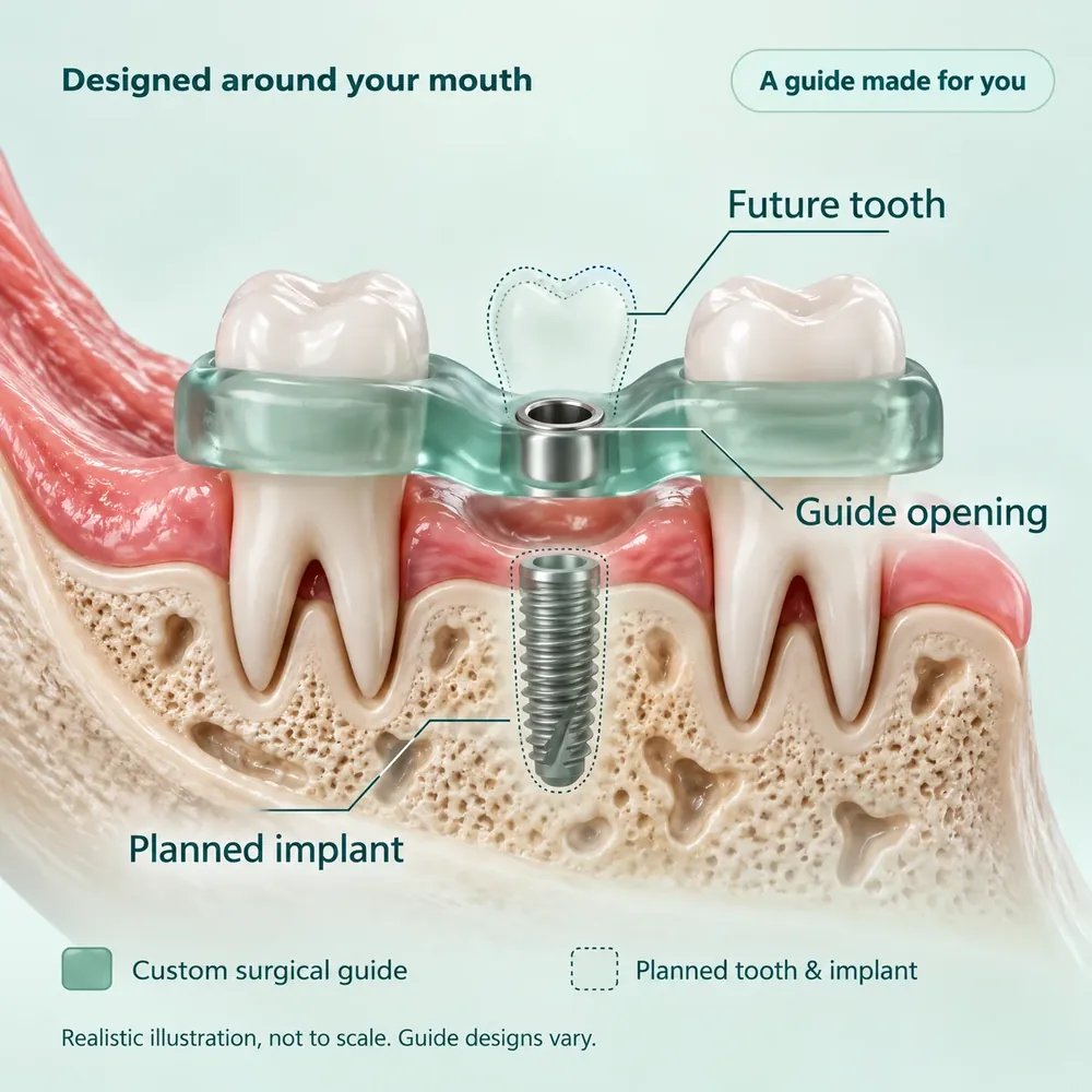
In suitable cases, yes. The 3D scan can be combined with a digital scan of your teeth, and the plan turned into a custom-made guide that rests on your teeth or gums and steers the drill to the planned position.3

Studies find that implants placed with a guide end up, on average, a little over one millimetre from the plan. That is why dentists still keep a safety margin of at least 2 mm around nerves.12 A guide is not needed for every case. Ask your dentist whether it would help in yours.

More about guided surgery, in guide 5

Does a 3D scan guarantee my implant will succeed?

No scan can promise that. Success also depends on your general health, whether you smoke, how you heal, and how well you clean and look after the implant.9 What the scan does is take a great deal of guesswork out of the planning.

Will I need more CBCT scans after treatment?

Only if there is a clinical reason, such as an implant that feels loose or a change in the feeling in your lip or chin. For routine checks on an implant that is causing no trouble, ordinary dental X-rays are usually enough.2

Can I have a copy of my scan?

Scans are stored in DICOM, the standard format for medical images, so other dentists and specialists can open them.4 Ask our team if you need a copy.

References

  1. U.S. Food and Drug Administration. Dental Cone-beam Computed Tomography. Content current as of 28 September 2020.

    fda.gov: Dental Cone-beam Computed Tomography
  2. Tyndall DA, Price JB, Tetradis S, Ganz SD, Hildebolt C, Scarfe WC. Position statement of the American Academy of Oral and Maxillofacial Radiology on selection criteria for the use of radiology in dental implantology with emphasis on cone beam computed tomography.

    Oral Surg Oral Med Oral Pathol Oral Radiol. 2012;113(6):817–826. Full text from the Academy (PDF). doi:10.1016/j.oooo.2012.03.005
  3. Jacobs R, Salmon B, Codari M, Hassan B, Bornstein MM. Cone beam computed tomography in implant dentistry: recommendations for clinical use.

    BMC Oral Health. 2018;18:88. Open access. doi:10.1186/s12903-018-0523-5
  4. Castellini (Cefla s.c.). iRYS imaging and planning software.

    castellini.com/en/software/irys
  5. American Dental Association. New ADA recommendations confirm dental imaging most effectively used in moderation.

    ADA News, 5 January 2026. adanews.ada.org. The recommendations, endorsed by the American Academy of Oral and Maxillofacial Radiology, are published in The Journal of the American Dental Association, January 2026: journal abstract
  6. Castellini (Cefla s.c.).

    X-Radius Trio, manufacturer’s brochure, 2014, hosted by a distributor: X-Radius Trio brochure (PDF). Castellini’s current imaging range: castellini.com/en/extraoral-devices
  7. RadiologyInfo.org, from the Radiological Society of North America and the American College of Radiology. Dental Cone Beam CT, patient information.

    radiologyinfo.org: Dental Cone Beam CT
  8. Ludlow JB, et al. Effective dose of dental CBCT: a meta analysis of published data and additional data for nine CBCT units.

    Dentomaxillofac Radiol. 2015;44(1):20140197. doi:10.1259/dmfr.20140197 (PubMed summary)
  9. U.S. Food and Drug Administration. Dental Implants: What You Should Know. Content current as of 29 October 2021.

    fda.gov: Dental Implants
  10. United Nations Scientific Committee on the Effects of Atomic Radiation (UNSCEAR). UNSCEAR 2000 Report to the General Assembly, press release, 6 June 2000. Worldwide average dose from natural radiation: 2.4 millisieverts a year.

    press.un.org
  11. American Dental Association. X-Rays/Radiographs. Oral Health Topics. Updated 26 March 2026.

    ada.org: X-Rays/Radiographs
  12. Tahmaseb A, Wu V, Wismeijer D, Coucke W, Evans C. The accuracy of static computer-aided implant surgery: a systematic review and meta-analysis.

    Clin Oral Implants Res. 2018;29(Suppl 16):416–435. doi:10.1111/clr.13346

This page gives general information and is not a substitute for advice from your own dentist, who will recommend what is right for you. Sources checked October 2026.

Back to top

التصوير ثلاثي الأبعاد في مركز ماجيك لطب وزراعة الأسنان

الصورة الكاملة قبل زراعة أسنانك

يجب أن تستقر زرعة الأسنان في عظم متين، بعيدًا عن الأعصاب والجيوب الأنفية. لذلك، وقبل وضع الزرعة، نلتقط صورة أشعة ثلاثية الأبعاد لفكّك تُسمّى تصوير CBCT، بجهاز Castellini X-Radius Trio في مركزنا. بعدها يستطيع طبيبك قياس العظم واختيار حجم الزرعة وموضعها على الشاشة أولًا.

  • بلا ألم
  • ينتهي في أقل من دقيقة
  • يُجرى هنا في مركزنا

ما يراه طبيب الأسنان

الصورة المسطّحة تبيّن ارتفاع العظم، لكنها لا تبيّن سماكته.

  • ارتفاع العظم. يمكن رؤيته.
  • قناة العصب. تُرى من الجانب فقط.
  • سماكة العظم. غير ظاهرة، ولذلك لا تكفي هذه الصورة وحدها لمعرفة هل يتّسع العظم للزرعة.
مقطع عرضي للفك السفلي من تصوير ثلاثي الأبعاد مقطع عرضي لعظم الفك تظهر فيه زرعة وسنّ جديد في أعلى العظم. سهم يبيّن أن ارتفاع العظم فوق قناة العصب 14.5 ملم، وخط يبيّن أن عرض العظم 6.8 ملم، وتظهر قناة العصب دائرةً داكنة في الأسفل. A B C D

الصورة ثلاثية الأبعاد يمكن تقطيعها إلى شرائح، فتظهر سماكة العظم وموضع العصب.

  • عرض العظم. قياسه هنا 6.8 ملم.
  • ارتفاع العظم. قياسه هنا 14.5 ملم فوق العصب.
  • قناة العصب. تحمل العصب إلى الشفة والذقن.
  • الزرعة والسنّ الجديد. بحجم وزاوية يُبقيانها بعيدة عن العصب.

اختر حرفًا لإبرازه في الصورة.

صور للتوضيح على سبيل المثال.

ما هو تصوير CBCT؟

CBCT هو اختصار للتصوير المقطعي المحوسب بالحزمة المخروطية. يدور ذراع الجهاز حول رأسك وهو يرسل حزمة مخروطية الشكل من الأشعة السينية عبر فكّك. ثم يحوّل الحاسوب ما التقطه إلى صورة ثلاثية الأبعاد لأسنانك وعظم فكّك والقنوات العظمية التي تمرّ فيها الأعصاب والجيوب الأنفية.1

التصوير سريع وغير مؤلم، ويُجرى هنا في المركز. يستخدمه أطباء الأسنان للتخطيط لزراعة الأسنان، وفي حالات أخرى لا تكفي فيها صورة الأشعة المسطّحة للإجابة عن السؤال.1

صورة الأشعة العادية تشبه الخريطة المسطّحة

ترى فيها مواقع الشوارع، لكنك لا تعرف ارتفاع المباني. كل شيء مضغوط في صورة واحدة.

تصوير CBCT يشبه مجسّمًا للكرة الأرضية تديره بيدك

يستطيع طبيبك تدويره وتكبيره وتصفّح فكّك شريحةً رقيقة بعد أخرى، ومن أي اتجاه.

لماذا يهمّ التصوير ثلاثي الأبعاد لزراعة أسنانك؟

الزرعة دعامة صغيرة من التيتانيوم تحلّ محلّ جذر السنّ المفقود. موضعها وطولها وزاوية ميلها تعتمد كلها على ما يوجد تحت اللثة.

  • يقيس العظم

    تحتاج الزرعة إلى عظم كافٍ يحيط بها من كل الجهات. يبيّن التصوير ارتفاع العظم وعرضه وشكله، فيعرف طبيبك قبل الجراحة هل يتّسع العظم للزرعة.23

  • يبيّن مسار العصب

    في الفك السفلي، يمرّ العصب المسؤول عن الإحساس في الشفة والذقن داخل قناة في العظم. رؤية هذه القناة بالأبعاد الثلاثة تساعد طبيبك على التخطيط لزرعة تبقى بعيدة عنها.23

  • يبيّن مدى قرب الجيب الأنفي

    فوق الأسنان الخلفية العلوية فراغات مملوءة بالهواء تُسمّى الجيوب الأنفية. يبيّن التصوير مقدار العظم الموجود تحتها.3

  • يتيح لطبيبك التجربة على الشاشة أولًا

    يستطيع طبيبك وضع زرعة افتراضية على الصورة وتجربة أحجام وأعماق وزوايا مختلفة، بما يناسب عظمك والسنّ الجديد الذي سيُركَّب فوقها.34

  • يبيّن الحاجة إلى عظم إضافي

    إذا كان العظم رقيقًا أو قليل الارتفاع، يساعد التصوير طبيبك على تحديد الحاجة إلى تطعيم عظمي أو رفع للجيب الأنفي قبل الزرعة أو معها.2

  • مفاجآت أقل يوم الجراحة

    بعض التفاصيل قد لا تُلاحَظ بسهولة في الصورة المسطّحة، مثل فتحة عصبية إضافية أو حافة عظمية ضيقة. واكتشافها في مرحلة التخطيط أفضل كثيرًا من اكتشافها أثناء الجراحة.3

توصي الأكاديمية الأمريكية لأشعة الفم والوجه والفكين بالتصوير المقطعي ثلاثي الأبعاد لأي موضع يُحتمل وضع زرعة فيه، وتعدّ تصوير CBCT الطريقة المفضّلة لذلك.2

وتصف توصيات جمعية طب الأسنان الأمريكية لعام 2026 تصوير CBCT بأنه التصوير المستخدم للتخطيط لجراحة الزراعة.5

جهازنا: Castellini X-Radius Trio

Castellini شركة إيطالية لتصنيع أجهزة طب الأسنان تأسست عام 1935. يلتقط جهاز X-Radius Trio لدينا صور الأشعة البانورامية العادية وصور CBCT ثلاثية الأبعاد في جهاز واحد. وهذا ما تعنيه مزاياه لك.6

يصوّر المنطقة المطلوبة فقط

اختر مساحة التصوير لترى ما تغطيه.

زرعة واحدة. يمكن حصر التصوير في الأسنان والعظم حول موضع الزرعة.

عدة زرعات في فك واحد. يغطي التصوير ذلك الفك كاملًا.

خطة للفم كله. صورة واحدة يمكن أن تشمل الفكّين معًا.

الإطار الأزرق هو المنطقة المصوَّرة، والإطار الأحمر يحدّد موضع الزرعة. كلما صغرت المساحة قلّ الجزء المعرَّض للأشعة من جسمك.12

  • تعرّض للأشعة بقدر الحاجة

    حزمة الأشعة نبضية، وللجهاز أنماط تصوير مختلفة، فيختار طبيبك الإعداد الذي يعطي التفاصيل الكافية لحالتك دون زيادة.

  • تحقّق سريع قبل التصوير

    مسند للرأس وأضواء توجيه تساعد على ضبط وضعيتك، وصورة أولية تؤكد أن التصوير موجَّه إلى المنطقة الصحيحة. وهذا يقلّل احتمال إعادة التصوير.

  • تفاصيل دقيقة

    تتكوّن الصورة ثلاثية الأبعاد من مكعبات صغيرة جدًا تشبه البكسلات في الصور الرقمية. في هذا الجهاز قد يصل حجمها إلى 0.075 ملم، أي أقل من عُشر المليمتر.

  • مفتوح وسريع

    تقف في جهاز مفتوح وليس داخل نفق، والتصوير نفسه يستغرق عادةً أقل من دقيقة.7

  • برنامج للتخطيط

    تُفتح الصورة في برنامج iRYS من Castellini، فيضع طبيبك زرعة افتراضية ويرى بُعدها عن قناة العصب ويحدّد مسافة أمان. وتُحفظ الصور بصيغة DICOM، وهي الصيغة المعتمدة للصور الطبية.4

مقارنة CBCT بأنواع التصوير الأخرى

لا يوجد نوع واحد من التصوير هو الأفضل لكل شيء، فلكل نوع سؤال يجيب عنه. اختر نوعًا لمقارنته بتصوير CBCT.

ما يضيفه تصوير CBCT

  • يبيّن سماكة العظم، وهو ما لا تستطيعه الصورة المسطّحة.23
  • يبيّن قناة العصب والجيب الأنفي بالأبعاد الثلاثة. أما الصورة المسطّحة فتُظهرهما من جهة واحدة فقط، فلا يمكن تقدير العمق.2
  • يتيح لطبيبك تخطيط موضع الزرعة على الشاشة.3

مزايا الأشعة العادية

  • جرعة الإشعاع فيها أقل.1
  • هي الخيار المناسب للفحوص الدورية وللتقييم الأولي قبل الزراعة.25
  • تكفي عادةً للمتابعة الدورية لزرعة لا تسبّب أي مشكلة.2

ما يقدّمه تصوير CBCT

  • مصمَّم للأسنان والفكّين، ويعطي تفاصيل العظم ثلاثية الأبعاد التي تحتاجها خطة الزراعة.3
  • جرعة الإشعاع فيه أقل عمومًا من التصوير الطبقي التقليدي.17
  • يُجرى في عيادة الأسنان وأنت واقف.

مزايا التصوير الطبقي في المستشفى

  • يُظهر الأنسجة الرخوة مثل العضلات والغدد بوضوح أكبر.7
  • يستخدمه الأطباء لحالات طبية تتجاوز الأسنان وعظم الفك.7

ما يقدّمه جهاز X-Radius Trio لدينا

  • مساحات تصوير متعددة، فيمكن حصر التصوير في المنطقة التي يحتاجها طبيبك.6
  • وسائل لضبط الوضعية وصورة أولية للتحقق، للحصول على صورة صحيحة من المرة الأولى.6
  • انتقال مباشر من الصورة إلى برنامج تخطيط الزراعة.4

من المفيد أن تعرف

  • أجهزة CBCT الحديثة الأخرى تقدّم مزايا مشابهة.
  • تختلف جرعة الإشعاع وجودة الصورة كثيرًا من جهاز إلى آخر، وبحسب مساحة التصوير والإعدادات المختارة.38
  • الأهم أن يكون التصوير مناسبًا لحالتك: أصغر مساحة وأقل جرعة تعطيان صورة واضحة بما يكفي.3

التصوير يدعم التخطيط الجيد، لكنه لا يغني عن الفحص السريري، ولا عن العلاج نفسه، ولا عن عنايتك بالزرعة بعد ذلك.9

الإشعاع والسلامة

يستخدم تصوير CBCT الأشعة السينية، ولذلك من حقك أن تسأل عن الإشعاع. إليك الصورة بوضوح.

أين يقع تصوير CBCT؟

  1. أشعة الأسنان العاديةأقل جرعة
  2. تصوير CBCT للأسنانفي الوسط
  3. التصوير الطبقي (CT)الأعلى عمومًا

يبيّن هذا الرسم الترتيب فقط، وارتفاع الأعمدة ليس بمقياس حقيقي.17

  • ما يعادل تقريبًا أسبوعين إلى شهر من الإشعاع الطبيعي اليومي

    وجدت مراجعة لقياسات أُجريت على أجهزة CBCT كثيرة ومختلفة أن متوسط الجرعة للبالغين نحو 84 ميكروسيفرت للصور الصغيرة ونحو 212 للصور الكبيرة.8 ويتلقى كل إنسان نحو 2,400 ميكروسيفرت سنويًا من مصادر طبيعية مثل الأرض والهواء والفضاء.10 هذه متوسطات لأجهزة متعددة وليست قياسًا لجهازنا، والجرعات تتفاوت كثيرًا بحسب الجهاز والإعدادات المستخدمة.

  • تصوير له سبب

    يفحصك طبيبك أولًا ويراجع صور الأشعة السابقة إن وُجدت. ولا يُجرى تصوير CBCT إلا إذا كانت المعلومات الإضافية ستفيد علاجك، وعندها تفوق فائدته الخطر الإشعاعي البسيط.111

  • أصغر مساحة تفي بالغرض

    تُحصر مساحة التصوير في أصغر حدّ ممكن يشمل ما يحتاج طبيبك إلى رؤيته.12

  • إعدادات تناسبك

    لا حاجة دائمًا إلى أعلى درجات الدقة. تُختار الإعدادات بحسب حجم جسمك وما يريد طبيبك معرفته.1

  • أخبرينا إذا كنتِ حاملًا أو تحتملين ذلك

    نرجو إخبارنا قبل أي تصوير بالأشعة، ليناقش طبيبك الخيارات معكِ.7

ماذا يحدث يوم التصوير؟

لا يحتاج التصوير إلى أي تحضير خاص.7

  1. إزالة الأغراض المعدنية

    سنطلب منك خلع النظارات والأقراط والقلائد ودبابيس الشعر وسمّاعات الأذن الطبية، وسنخبرك إن كان عليك نزع أطقم الأسنان أو أي تركيبات سنّية متحركة أخرى.7

  2. اتخاذ الوضعية

    تقف داخل الجهاز. مسند للرأس يثبّتك، وأضواء توجيه تساعدنا على ضبط وضعيتك.6

  3. الثبات أثناء التصوير

    يدور الذراع حول رأسك. ابقَ ثابتًا وتنفّس بشكل طبيعي. لن تشعر بشيء.7

  4. متابعة يومك

    لا تحتاج إلى فترة نقاهة. يدرس طبيبك الصور ثلاثية الأبعاد ويضع خطة الزرعة ثم يشرحها لك.7

أسئلة يطرحها المرضى

هل أحتاج إلى تصوير CBCT لزراعة الأسنان؟

توصي الإرشادات المهنية بالتصوير ثلاثي الأبعاد عند التخطيط لجراحة الزراعة.25 يفحص طبيبك فمك أولًا ويراجع صور الأشعة السابقة، ثم يشرح لك لماذا يناسب التصوير حالتك.

إذا أجريت تصوير CBCT في مكان آخر مؤخرًا، فأخبرنا أو أحضر الصورة معك، فقد لا تحتاج إلى تصوير جديد.111

لماذا لا تكفي صورة الأشعة العادية؟

صورة الأشعة العادية تضغط الفك في صورة واحدة مسطّحة، فلا تبيّن سماكة العظم، ولا هل العصب أقرب إلى جهة الخد أم إلى جهة اللسان. والزرعة جسم ثلاثي الأبعاد يوضع في عظم ثلاثي الأبعاد، ولهذا توصي الإرشادات بالتصوير ثلاثي الأبعاد للتخطيط لها.2

وما زلنا نستخدم الأشعة العادية للفحوص الدورية والتقييم الأولي، لأن جرعة الإشعاع فيها أقل.1

هل التصوير مؤلم؟ وهل له آثار جانبية؟

لا، ليس مؤلمًا. لا توجد حقن، ولا يلامسك شيء سوى المساند التي تثبّت رأسك. والأشعة السينية لا تبقى في جسمك، ولا توجد آثار جانبية فورية، ويمكنك العودة إلى نشاطك مباشرة.7

وكأي تصوير بالأشعة، ينطوي هذا التصوير على جرعة صغيرة من الإشعاع. انظر قسم الإشعاع والسلامة.

كم يستغرق التصوير؟

يستغرق التصوير نفسه عادةً أقل من دقيقة: بضع ثوانٍ للمساحة الصغيرة، وحتى نحو 40 ثانية لتصوير الفم كاملًا.7 خصّص بضع دقائق إضافية لخلع الحليّ واتخاذ الوضعية.

هل هو آمن؟ وما مقدار الإشعاع الذي سأتعرّض له؟

تصوير CBCT ليس خاليًا من الإشعاع. جرعته أعلى من صورة أشعة الأسنان العادية وأقل عمومًا من التصوير الطبقي في المستشفى.1 وفي أجهزة CBCT المختلفة، يعادل متوسط الجرعة تقريبًا ما يتلقاه أي إنسان من الإشعاع الطبيعي في أسبوعين إلى شهر.810

وعندما يكون التصوير ضروريًا لعلاجك، تفوق فائدته هذا الخطر البسيط.1 ويعتمد المقدار الدقيق على مساحة التصوير والإعداد المستخدم، ولهذا نختار أصغر مساحة وأقل إعداد يفيان بالغرض.

هل هو نفسه التصوير الطبقي في المستشفى؟

هو تقنية قريبة منه تعطي صورًا ثلاثية الأبعاد أيضًا، لكنها مصمَّمة للأسنان والفكّين. وجرعة الإشعاع في تصوير CBCT للأسنان أقل عمومًا من التصوير الطبقي التقليدي. أما التصوير الطبقي في المستشفى فأفضل في إظهار الأنسجة الرخوة مثل العضلات والغدد، ولذلك يستخدمه الأطباء لأغراض أخرى.17

بماذا أخبركم قبل التصوير؟

في حال وجود حمل أو احتمال وجوده، نرجو إخبارنا ليناقش الطبيب إجراء التصوير أو تأجيله.7 ونرجو إحضار ما يتوفر عن أي صور أشعة حديثة للأسنان.11 وسنخبرك بالحليّ والتركيبات السنّية المتحركة التي يجب خلعها.

لديّ حشوات أو تيجان أو زرعات أخرى. هل تؤثر في الصورة؟

قد يسبّب المعدن خطوطًا لامعة في صورة CBCT. يأخذ طبيبك ذلك في الحسبان عند قراءة الصورة، وتستطيع برامج التصوير تخفيف هذه الخطوط.3

هل يُستخدم التصوير في الزراعة الموجَّهة؟

نعم، في الحالات المناسبة. يمكن دمج الصورة ثلاثية الأبعاد مع مسح رقمي لأسنانك، وتحويل الخطة إلى دليل جراحي يُصنع خصيصًا لك، يستقر على أسنانك أو لثتك ويوجّه أداة الحفر إلى الموضع المخطط له.3

تبيّن الدراسات أن الزرعات الموضوعة باستخدام الدليل تبتعد عن الخطة بما يزيد قليلًا على مليمتر واحد في المتوسط. ولهذا يُبقي أطباء الأسنان هامش أمان لا يقل عن 2 ملم حول الأعصاب.12 ولا تحتاج كل الحالات إلى دليل، فاسأل طبيبك إن كان مفيدًا لحالتك.

المزيد عن الجراحة الموجَّهة في الدليل 5

هل يضمن التصوير ثلاثي الأبعاد نجاح الزرعة؟

لا يوجد تصوير يضمن ذلك. فالنجاح يعتمد أيضًا على صحتك العامة، وعلى التدخين، وعلى سرعة الالتئام، وعلى عنايتك بتنظيف الزرعة والمحافظة عليها.9 لكن التصوير يزيل قدرًا كبيرًا من التخمين في مرحلة التخطيط.

هل أحتاج إلى تصوير CBCT آخر بعد العلاج؟

فقط إذا وُجد سبب سريري، مثل زرعة تشعر بأنها تتحرك أو تغيّر في الإحساس في الشفة أو الذقن. أما المتابعة الدورية لزرعة لا تسبّب أي مشكلة فتكفيها عادةً صور الأشعة العادية.2

هل أستطيع الحصول على نسخة من صورتي؟

تُحفظ الصور بصيغة DICOM، وهي الصيغة المعتمدة للصور الطبية، فيستطيع أطباء الأسنان والاختصاصيون الآخرون فتحها.4 اطلب نسخة من فريقنا إن احتجت إليها.

المراجع

المراجع مدرجة بلغتها الأصلية، الإنجليزية.

  1. U.S. Food and Drug Administration. Dental Cone-beam Computed Tomography. Content current as of 28 September 2020.

    fda.gov: Dental Cone-beam Computed Tomography
  2. Tyndall DA, Price JB, Tetradis S, Ganz SD, Hildebolt C, Scarfe WC. Position statement of the American Academy of Oral and Maxillofacial Radiology on selection criteria for the use of radiology in dental implantology with emphasis on cone beam computed tomography.

    Oral Surg Oral Med Oral Pathol Oral Radiol. 2012;113(6):817–826. Full text from the Academy (PDF). doi:10.1016/j.oooo.2012.03.005
  3. Jacobs R, Salmon B, Codari M, Hassan B, Bornstein MM. Cone beam computed tomography in implant dentistry: recommendations for clinical use.

    BMC Oral Health. 2018;18:88. Open access. doi:10.1186/s12903-018-0523-5
  4. Castellini (Cefla s.c.). iRYS imaging and planning software.

    castellini.com/en/software/irys
  5. American Dental Association. New ADA recommendations confirm dental imaging most effectively used in moderation.

    ADA News, 5 January 2026. adanews.ada.org. The recommendations, endorsed by the American Academy of Oral and Maxillofacial Radiology, are published in The Journal of the American Dental Association, January 2026: journal abstract
  6. Castellini (Cefla s.c.).

    X-Radius Trio, manufacturer’s brochure, 2014, hosted by a distributor: X-Radius Trio brochure (PDF). Castellini’s current imaging range: castellini.com/en/extraoral-devices
  7. RadiologyInfo.org, from the Radiological Society of North America and the American College of Radiology. Dental Cone Beam CT, patient information.

    radiologyinfo.org: Dental Cone Beam CT
  8. Ludlow JB, et al. Effective dose of dental CBCT: a meta analysis of published data and additional data for nine CBCT units.

    Dentomaxillofac Radiol. 2015;44(1):20140197. doi:10.1259/dmfr.20140197 (PubMed summary)
  9. U.S. Food and Drug Administration. Dental Implants: What You Should Know. Content current as of 29 October 2021.

    fda.gov: Dental Implants
  10. United Nations Scientific Committee on the Effects of Atomic Radiation (UNSCEAR). UNSCEAR 2000 Report to the General Assembly, press release, 6 June 2000. Worldwide average dose from natural radiation: 2.4 millisieverts a year.

    press.un.org
  11. American Dental Association. X-Rays/Radiographs. Oral Health Topics. Updated 26 March 2026.

    ada.org: X-Rays/Radiographs
  12. Tahmaseb A, Wu V, Wismeijer D, Coucke W, Evans C. The accuracy of static computer-aided implant surgery: a systematic review and meta-analysis.

    Clin Oral Implants Res. 2018;29(Suppl 16):416–435. doi:10.1111/clr.13346

تقدّم هذه الصفحة معلومات عامة ولا تغني عن استشارة طبيب أسنانك، فهو من يحدّد ما يناسب حالتك. تم التحقق من المصادر في تشرين الأول (أكتوبر) 2026.

العودة إلى الأعلى

Why we use Straumann® implants

At Magic Tooth Dental Center we want your new tooth to feel secure, look natural and last for many years. That is why we use Straumann, a Swiss implant maker trusted by dentists around the world.

  • A Swiss company, founded in 1954
  • Studied in patients for 10 years and more
  • Original Straumann parts throughout
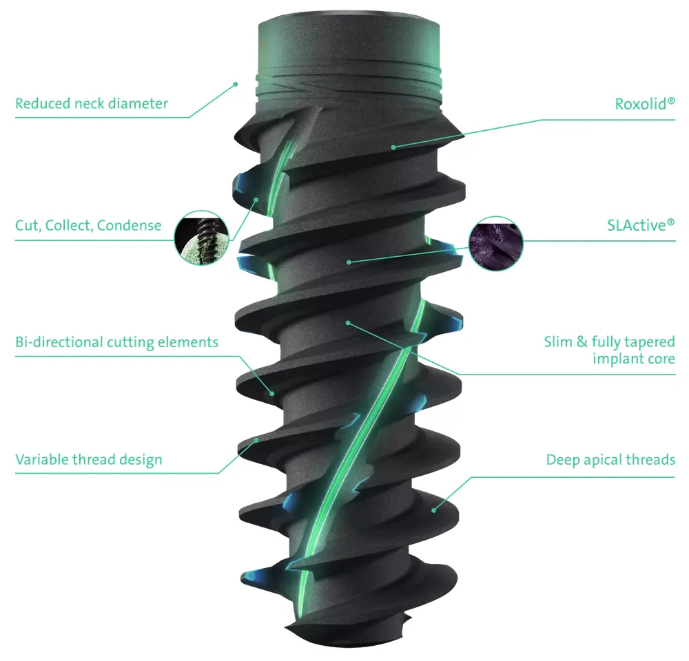
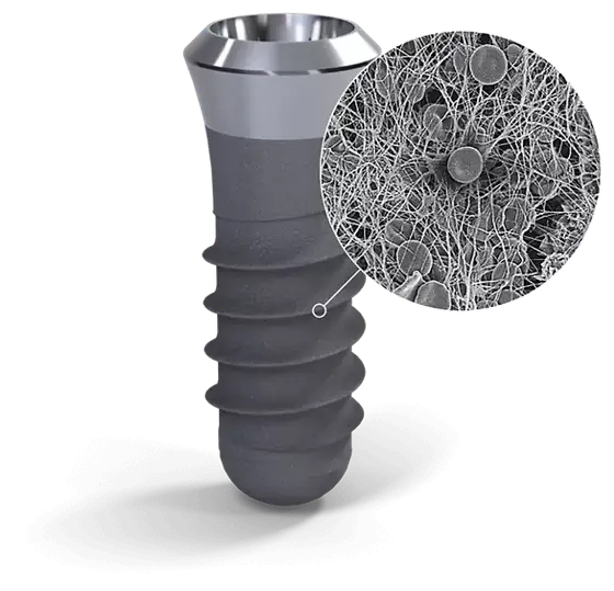
A Straumann dental implant. A magnified circle shows fine blood fibres and cells covering its surface.
A Straumann implant. The circle shows its surface under a microscope, with blood fibres and cells settling on it as healing begins.
Straumann

We use genuine Straumann implants and original parts, from the start of your treatment to the finish.

How a dental implant works

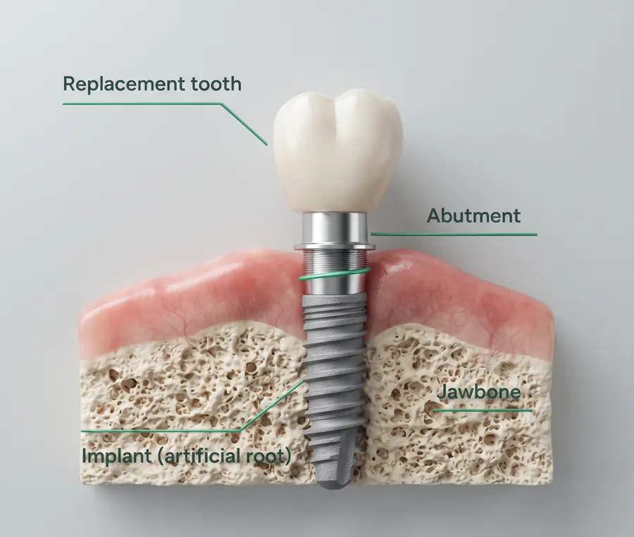
A dental implant is a small screw that takes the place of a missing tooth’s root. A connector called an abutment sits on top of it and holds your new crown, bridge or denture.

Over the following weeks your jawbone grows onto the implant and holds it firmly, the way it would hold a natural root. The better that bond, the stronger and longer-lasting your new tooth. This is where Straumann technology helps.

A word you may hear: osseointegration (oss-ee-oh-in-teh-gray-shun)It simply means your bone growing onto the implant and holding it in place.

Cross-section of a jaw. An implant, labelled as an artificial root, sits in the jawbone below the gum. An abutment on top of it holds the replacement tooth.
Simplified illustration, not a specific Straumann model.

Your treatment, step by step

  1. Check-up and planning

    We examine your teeth, gums and bone, talk about what you would like, and plan the right implant for you.

  2. Placing the implant

    The implant is placed in your jawbone. In suitable cases this can be done on the same day a tooth is removed.

  3. Healing

    Your bone grows onto the implant surface. Straumann surfaces are designed to make this reliable and, with SLActive, quicker.

  4. Your new tooth

    A crown, bridge or denture is fixed onto the implant and made to match your natural teeth.

Why Straumann

There are hundreds of implant brands. We chose Straumann for its Swiss engineering, the technology it developed itself, and some of the most thorough long-term research in dentistry.

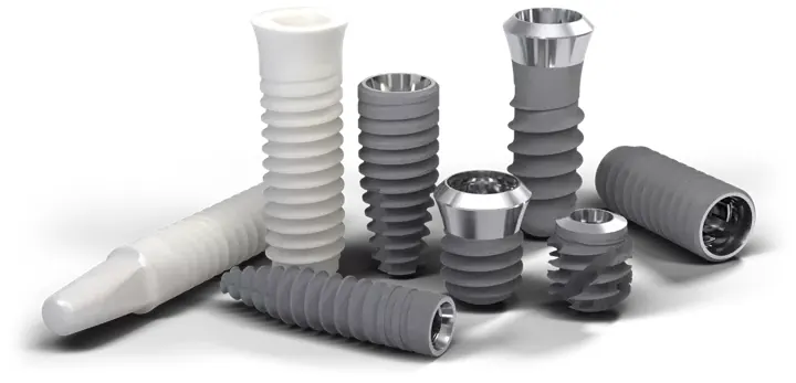
A range of Straumann implants in different shapes and sizes, in grey titanium and white ceramic.
  • Swiss roots

    Straumann was founded in Switzerland in 19549 and has made dental implants since the 1970s.

  • Decades of research

    Its implants are among the most studied in dentistry, with patients followed for 10 years and more.2, 6

  • Its own inventions

    The SLA® and SLActive® surfaces and the Roxolid® material were all developed by Straumann.

  • Known around the world

    Dentists in many countries work with Straumann, so your implant can be looked after wherever life takes you.

What the studies and figures show

98.8%

of 511 SLA implants were still in place after 10 years, in a University of Bern study.2

98.2%

of SLActive implants that were given a temporary tooth on the day of surgery were still in place after 10 years.6

over35%

of the world’s dental implant market is held by the Straumann Group, by its own 2025 estimate.9

Study results describe groups of patients and cannot promise an individual result. Market share shows how widely the Straumann Group’s implant brands are used. It is not a ranking of quality.

The technology, in plain words

The names can be confusing, so here is the short version. Every implant has a surface, a material and a shape. Straumann has its own technology for each one.

The surface
What your bone grows onto. SLA or SLActive.
The material
What the implant is made of. Roxolid.
The shape
How the implant grips your bone. BLX.
Surface

SLA®: the proven classic

SLA is the surface that made Straumann famous. The name stands for Sandblasted, Large-grit, Acid-etched. In simple terms, the implant is given a very fine, rough texture, far too small to see. Bone holds on to a rough surface better than a smooth one, so it grows into the texture and locks the implant in place.

What this means for you

  • A strong, lasting bond. In a 10-year study at the University of Bern, 98.8% of 511 SLA implants were still in place.2
  • A long track record. SLA has been used in patients for more than 25 years.1
  • Few problems over time. In the same study, fewer than 2 in 100 implants developed an infection around the implant over 10 years.2
  • Reliable healing. The implant usually bonds with the bone in about 6 to 8 weeks.5
Three Straumann implants with a fine, matt grey surface.
Straumann implants. The matt grey finish is the roughened surface.
Surface

SLActive®: faster early healing

SLActive keeps the proven SLA texture and adds one upgrade: the surface is “hydrophilic”, which simply means water-loving. The implant is kept in a protective liquid until the moment it is placed. When it touches your blood, the surface soaks it up straight away, so the cells that build bone can get to work sooner.

What this means for you

  • Shorter early healing. About 3 to 4 weeks in suitable cases, instead of 6 to 8 weeks with SLA.5
  • More support in the first weeks. In a study in people, new bone attached to SLActive faster than to SLA during the first four weeks. By six weeks the two were level.3
  • Your new tooth sooner. In a 10-year study, 98.2% of SLActive implants given a temporary tooth on the day of surgery were still in place, and 97.1% of those given one about a month later.6
  • Help when healing is harder. In a small two-year study, SLActive implants did as well in people with well-controlled type 2 diabetes as in people without diabetes.11
Standard SLAAt first, a drop of blood sits on top.
SLActiveThe blood spreads across the surface at once.
Simplified drawing, not to scale.
Material

Roxolid®: a stronger metal

Most dental implants are made of pure titanium. Straumann developed its own blend, called Roxolid, made specifically for dental implants. It is about 85% titanium and 15% zirconium.8

What this means for you

  • Extra strength. Roxolid is stronger than pure titanium.8
  • A smaller implant can do the job. Narrower or shorter implants can be used where bone is thin or the gap between teeth is tight.8
  • Gentler treatment. In some cases a smaller implant means a bone graft is not needed. That is less surgery and less recovery time.8
  • Kind to your bone. Roxolid bonds with bone very well, just as titanium does.8
  • Titanium, about 85%
  • Zirconium, about 15%
What Roxolid is made of. Every BLX implant is made from Roxolid.
Shape

BLX: a firm grip from the first moment

BLX is the name of an implant shape. It narrows towards the tip and has deep threads that hold tightly in the bone as soon as the implant is placed. That early grip is what makes immediate treatment possible: placing the implant on the same day a tooth is removed and, when conditions allow, fitting a temporary tooth straight away.

What this means for you

  • A firm hold from day one, even where bone is naturally softer.7
  • Fewer visits, less waiting. In suitable cases the tooth can be removed and the implant placed in one appointment.
  • Strong and slim. Every BLX implant is made from Roxolid.4
  • Gentle on the bone. The slim top is designed to put less stress on the bone around your new tooth.7
  • Planned in advance. BLX works with Straumann’s digital planning tools, which help your dentist decide the implant’s position before treatment.7

See the BLX shape up close

Three Straumann BLX implants with wide, deep threads.
Straumann BLX implants, with their deep threads.
All three together

BLX SLActive®: firm on day one, faster healing after

This is Straumann’s most advanced combination. It brings the BLX shape, the Roxolid material and the SLActive surface together in one implant.

What this means for you

  • Support from the first day and through healing. The BLX shape gives the early grip, and SLActive helps bone attach sooner.3, 7
  • A same-day tooth, for some patients. If your bone and gums are suitable, you can have an implant and a temporary tooth on the same day.7
  • When your dentist may suggest it. When you would like teeth as soon as possible, when your bone is softer, or when healing may be slower.
Shape
BLXA firm grip on day one
Material
RoxolidStrong, so it can be slim
Surface
SLActiveFaster early healing
Together
BLX SLActiveAll three in one implant

How long does early healing take?

Weeks for the implant to bond with your bone, in suitable cases.5

SLA6 to 8 weeks
SLActive3 to 4 weeks
These are Straumann’s guideline times for early healing. They are not a promise of complete healing, or of the date your final crown will be fitted. Your dentist decides the timing by checking how you heal.5, 6

BLX up close: every detail has a job

The BLX implant is shaped to hold firmly from the very first moment. Choose a number to see what each part does.

A Straumann BLX implant with eight labelled design features. Each one is explained in the list that follows.

Tap a number on the picture, or a line in the list.

  1. The top of the implant is slimmer, which is designed to put less stress on the bone around it. This helps protect the bone and gum that frame your new tooth.

  2. The titanium and zirconium blend is stronger than pure titanium, so a slimmer implant can still take the load of chewing.

  3. As the implant goes in, a groove along its side collects the tiny chips of bone that are cut away and presses them back around the implant.

  4. The surface draws in blood straight away, which helps bone attach sooner during the first, most delicate weeks of healing.

  5. The cutting edges work in both directions, so your dentist can adjust the implant’s position precisely while placing it.

  6. A narrow core that tapers to the tip needs only a small opening in the jaw, which keeps more of your own bone.

  7. The threads are thin and change along the length of the implant. They are designed to give a firm first hold in every type of bone.

  8. Deep threads at the tip anchor into the bone. This strong first hold is what allows immediate treatment.

Feature names and descriptions are based on Straumann’s BLX product information.7

Which option might suit you?

Your dentist recommends the implant that fits your bone, your health and your goals. This guide shows what you are likely to hear about, so you can come with good questions.

What matters most to you?

A guide to help you ask questions. It is not a treatment plan.

If you would like your new tooth as soon as possible

Your dentist may talk to you about

BLX SLActive

The BLX shape grips firmly from the first moment, and the SLActive surface shortens early healing to about 3 to 4 weeks in suitable cases. Some patients can leave with a temporary tooth on the same day.5, 7

Surface
SLActive
Material
Roxolid
Early healing
About 3 to 4 weeks

Read about BLX SLActive

If you want the longest track record

Your dentist may talk to you about

SLA

SLA is Straumann’s classic surface. It has been used in patients for more than 25 years, and in a 10-year study 98.8% of 511 implants were still in place.1, 2

Surface
SLA
Material
Titanium or Roxolid
Early healing
About 6 to 8 weeks

Read about SLA

If you are having a tooth taken out

Your dentist may talk to you about

BLX or BLX SLActive

BLX was designed for immediate treatment. In suitable cases the implant can be placed in the same visit as the tooth removal, which means fewer appointments and less waiting.7

Surface
SLA or SLActive
Material
Roxolid
Early healing
Depends on the surface

Read about BLX

If you have been told your bone is thin or soft

Your dentist may talk to you about

Roxolid, often in the BLX shape

Roxolid is stronger than pure titanium, so a narrower or shorter implant can do the job, and in some cases a bone graft is not needed. The BLX shape is designed to hold well even in softer bone.7, 8

Surface
SLA or SLActive
Material
Roxolid
Early healing
Depends on the surface

Read about Roxolid

If you heal slowly or have diabetes

Your dentist may talk to you about

SLActive

SLActive is designed to get healing started sooner. In a small two-year study, SLActive implants did as well in people with well-controlled type 2 diabetes as in people without diabetes. Your dentist will look at your general health first.11

Surface
SLActive
Material
Titanium or Roxolid
Early healing
About 3 to 4 weeks

Read about SLActive

The four options side by side

Highlighted rows match your choice above.

The four Straumann implant options compared
OptionSurfaceMaterialEarly healingMain advantage for you
SLAclassic shapesSLATitanium or RoxolidAbout 6 to 8 weeksProven long-term results
SLActiveclassic shapesSLActiveTitanium or RoxolidAbout 3 to 4 weeksFaster early healing, earlier teeth
BLXBLX shapeSLARoxolidAbout 6 to 8 weeksSame-day placement with a firm grip
BLX SLActiveBLX shapeSLActiveRoxolidAbout 3 to 4 weeksSame-day placement plus faster healing

Healing times are for suitable cases. Same-day placement depends on your bone and gums.4, 5

Why not a cheaper implant?

Many implant brands are available, at very different prices. This is what sets Straumann apart from many of them.

  • More long-term evidence

    Few implants have been followed as closely, or for as long, in published studies. Many lower-cost brands have little long-term data.2, 6

  • A faster-healing surface

    SLActive is Straumann’s own technology. It roughly halves early healing time compared with Straumann’s standard SLA surface.5

  • Fewer bone grafts, in some cases

    Because Roxolid is so strong, a narrower implant can sometimes be used. That may spare you a bone graft.8

  • Care wherever you live

    If you move abroad and need a small adjustment years from now, dentists in many countries know Straumann and can get the right parts. Some budget brands disappear from the market.

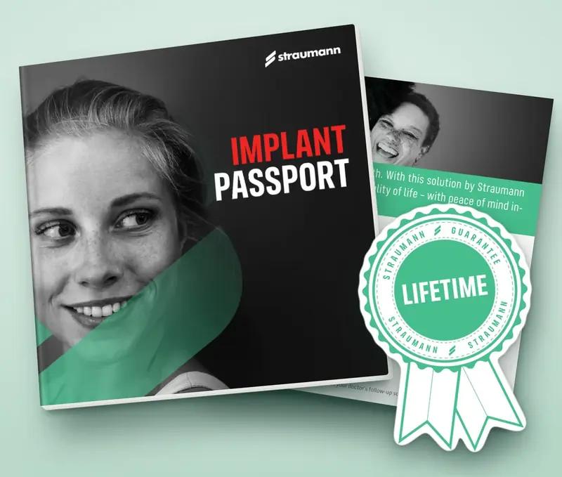
Straumann lifetime guarantee seal.

Backed by a lifetime guarantee

Straumann guarantees its implants for life, should one ever need to be replaced. Conditions apply.12

See what comes with your implant

An honest note. No implant can promise success by itself. Your health, careful planning and good home care all play a part. The points above are specific advantages of the technology. They do not mean one implant is the best choice for everyone.3, 10

Your new teeth come with a passport

Every Straumann implant we place is recorded in your own Implant Passport. It is yours to keep.

  • A lifetime guarantee on the implant

    Straumann guarantees its implants for life, should one ever need to be replaced. The guarantee is arranged through your dentist. Conditions apply, including the use of original Straumann parts, and terms can differ by country.12

  • Every part on record

    The original label of each part goes into your booklet, so any dentist can see exactly which implant you have. Bring it to every check-up.

  • Original parts, start to finish

    We use original Straumann parts throughout your treatment. Each one is made to fit the implant precisely, and replacement parts stay available for many years.

  • Known in more than 100 countries

    Straumann products are available in more than 100 countries, so help is nearby wherever life takes you.13

A Straumann Implant Passport booklet, shown with a green Straumann lifetime guarantee seal.

Questions patients ask us

Every smile is different. These are the questions we hear most often. Tap one to see the answer.

What is the difference between SLA, SLActive, Roxolid and BLX?

They describe different parts of the same implant. SLA and SLActive are surfaces: SLA is the proven classic, and SLActive is the faster-healing version. Roxolid is the material the implant is made from. BLX is the implant’s shape. One implant can combine them, for example a BLX implant made of Roxolid with an SLActive surface.4

Can I have a new tooth on the same day?

Sometimes. If your bone and gums are suitable, an implant and a temporary tooth can be placed on the same day, especially with BLX. Your dentist needs to examine you first. Your final tooth is made and fitted later, once healing is complete.6, 7

Will my implant be fully healed in three to four weeks?

Not necessarily. Three to four weeks is Straumann’s guideline for early healing with SLActive in suitable cases. It is not a promise that healing is complete, or that your final crown will be ready by then. Your dentist checks how you are healing and decides the timing.5, 6

Will I still need a bone graft?

It depends on your bone. Because Roxolid is strong, a narrower implant can sometimes be used, which may mean a graft is not needed. Your dentist can tell you after examining your jaw.8

Is SLActive suitable if I have diabetes or heal slowly?

It is often a good choice. In a small two-year study, SLActive implants made of Roxolid did as well in people with well-controlled type 2 diabetes as in people without diabetes. Your dentist will review your general health before recommending anything.11

How long does a Straumann implant last?

Implants are made to last for many years. In long-term studies, 97% to 99% of Straumann implants were still in place after 10 years. How long yours lasts also depends on your health, on cleaning well every day and on regular check-ups.2, 6

Why not choose a cheaper implant?

Price matters, but so does proof. Straumann implants have been followed in published studies for 10 years and more, the parts stay available, and the implant carries a lifetime guarantee. With a little-known brand, long-term evidence and spare parts can be harder to find.2, 12

What if I move abroad years from now?

Straumann is used by dentists around the world, and your implant passport records exactly which implant you have. That makes it easier for another dentist to find the right parts if you ever need an adjustment.

Which option is right for me?

That depends on your bone, your gums, your health and how soon you would like your new tooth. At your consultation we look at all of these and recommend the implant that suits you best.

Sources and references

The small numbers on this page link to these sources. Each opens in a new tab. Links checked on 10 October 2026.

  1. Straumann.

    The SLA surface and its clinical evidence.

    Manufacturer information. SLA was introduced in 1998.
  2. Buser D, Janner SFM, Wittneben JG, Brägger U, Ramseier CA, Salvi GE (2012).

    10-year survival and success rates of 511 titanium implants with a sandblasted and acid-etched surface.

    Clinical Implant Dentistry and Related Research 14(6): 839–851. 98.8% of implants survived; 1.8% developed peri-implantitis over 10 years.
  3. Lang NP, Salvi GE, Huynh-Ba G, Ivanovski S, Donos N, Bosshardt DD (2011).

    Early osseointegration to hydrophilic and hydrophobic implant surfaces in humans.

    Clinical Oral Implants Research 22(4): 349–356. More bone contact with SLActive in the first four weeks; no difference by six weeks.
  4. Straumann.

    BLX Implant System overview (PDF).

    Lists BLX implants in Roxolid with SLA and SLActive surfaces. Availability varies by country.
  5. Straumann.

    SLActive product information (PDF).

    Manufacturer guidance: healing time reduced from 6–8 weeks to 3–4 weeks.
  6. Nicolau P, Guerra F, Reis R, Krafft T, Benz K, Jackowski J (2019).

    10-year outcomes with immediate and early loaded implants with a chemically modified SLA surface.

    Quintessence International 50(2): 114–124. Survival of SLActive implants: 98.2% with a temporary tooth fitted on the day of surgery, 97.1% with one fitted about a month later. This study does not cover the newer BLX shape.
  7. Straumann.

    BLX Implant System brochure (PDF).

    Manufacturer description of the BLX design features.
  8. Straumann.

    Implant materials and Roxolid.

    Manufacturer information: 85% titanium, 15% zirconium.
  9. Straumann Group.

    Annual Report 2025 (PDF).

    Company history, and the company’s own estimate of an implant market share above 35%, across all of the Group’s brands.
  10. Vílchez B, Caneiro L, Lima C, Sanz-Sánchez I, Montero E, Figuero E, Blanco J, Sanz M (2026).

    Clinical and radiographic performance of two distinct sandblasted, large-grit, acid-etched implant surfaces: a split-mouth randomized clinical trial.

    Clinical Oral Implants Research 37(2): 141–154. A head-to-head trial of two surfaces of this type, listed for balance.
  11. Cabrera-Domínguez JJ, Castellanos-Cosano L, Torres-Lagares D, Pérez-Fierro M, Machuca-Portillo G (2020).

    Clinical performance of titanium-zirconium implants with a hydrophilic surface in patients with controlled type 2 diabetes mellitus: 2-year results.

    Clinical Oral Investigations 24(7): 2477–2486. 28 patients; no implants lost in either group.
  12. Straumann.

    The Straumann Guarantee.

    Terms of the implant guarantee. It is addressed to the treating dentist and varies by country.
  13. Straumann.

    The Straumann Group.

    Company overview: products and services available in more than 100 countries.

Rebuilding jawbone for a dental implant

A dental implant needs enough healthy bone to hold it. When bone has been lost, we can rebuild it with two materials from botiss, a German manufacturer: a bone graft called cerabone® +HyA and a protective cover called Jason® membrane. This page explains what they are, how they work and how they compare with other options.

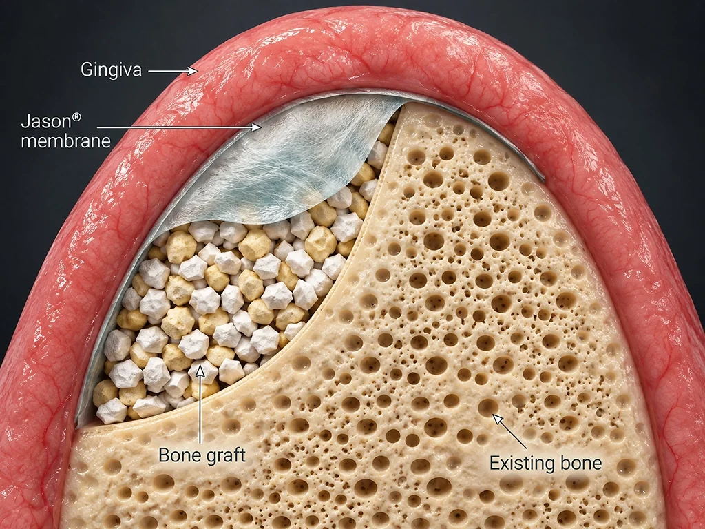
A look inside the repair
Cross-section of a jaw ridge where bone is being rebuilt. Pink gum, labelled gingiva, arches over the top. Under it a thin white sheet, the Jason membrane, covers a space packed with small bone graft granules, which rest against the sponge-like existing bone.
  1. Explore the picturePoint to any part with your mouse, tap it, or choose one of the four buttons, to see what it is and what it does.
  2. GumDentists call it the gingiva. Once the graft and the membrane are in place, your gum is closed over them. Gum grows much faster than bone, which is why the membrane sits between the two.
  3. Jason® membraneA very thin sheet of natural collagen, laid over the graft like a blanket. It keeps fast-growing gum out of the space while new bone forms. Your body absorbs it as you heal.
  4. Bone graft: cerabone® +HyAThe space where bone was lost is filled with this soft, mouldable graft and shaped to the contour your jaw needs. Over the following months your own bone grows through it.
  5. Your own boneThe jawbone you already have, labelled “existing bone” in the picture. After a tooth is lost, the bone around it often shrinks. An implant needs enough bone to hold it firmly.

Two materials, two jobs

Bone grows slowly. Gum grows quickly. Left alone, gum would fill an empty space long before bone could. So the graft gives new bone something to grow on, and the membrane keeps gum out while that happens. Dentists call this guided bone regeneration.5

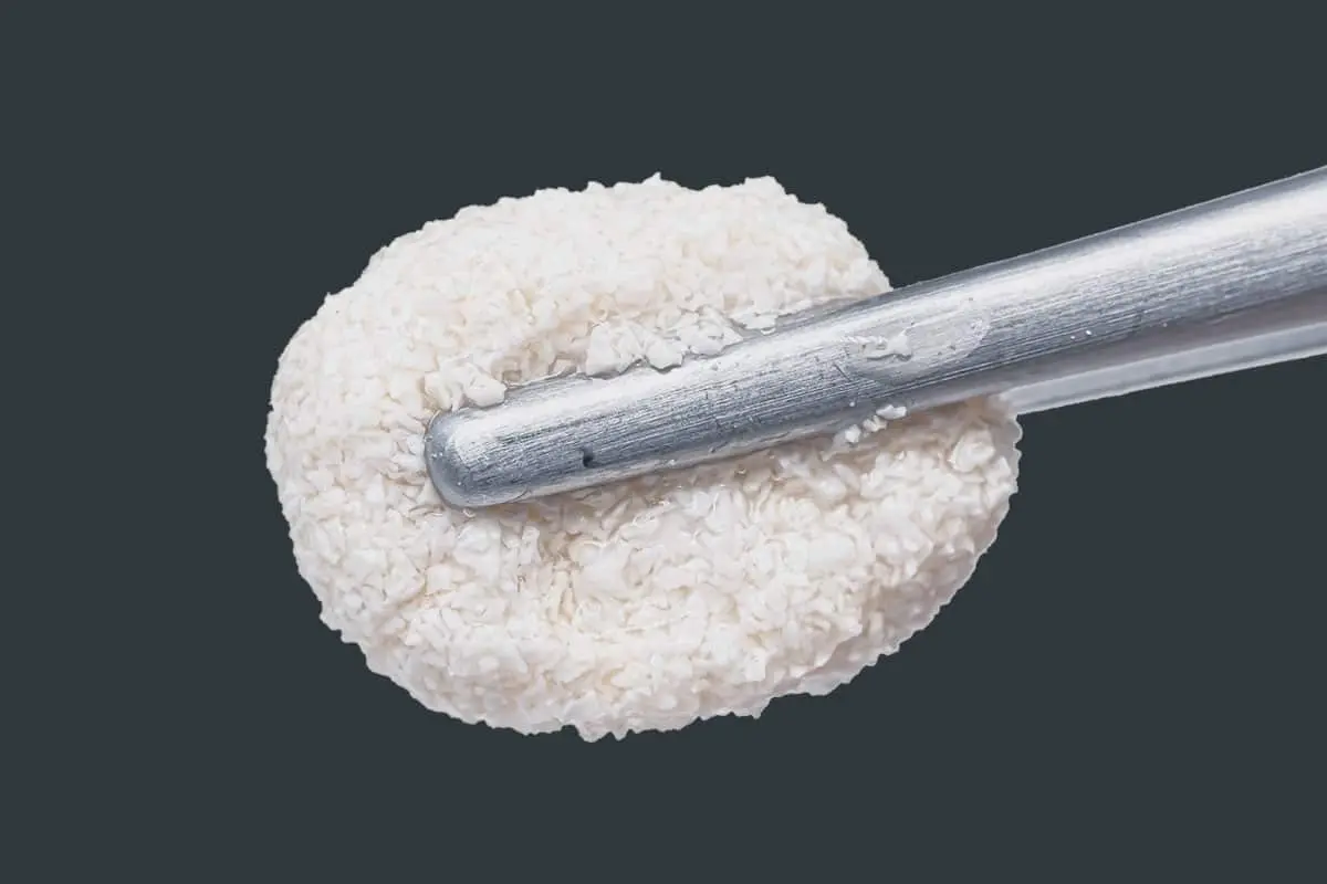
A soft ball of moistened cerabone +HyA granules holding together on the tip of a dental instrument.
The graft after mixing. The granules hold together instead of scattering.

cerabone® +HyA

Bone graft with hyaluronic acid

Small granules of natural bone mineral, mixed with hyaluronic acid. A few drops of sterile liquid turn them into a soft, mouldable material that dentists often call “sticky bone”.1

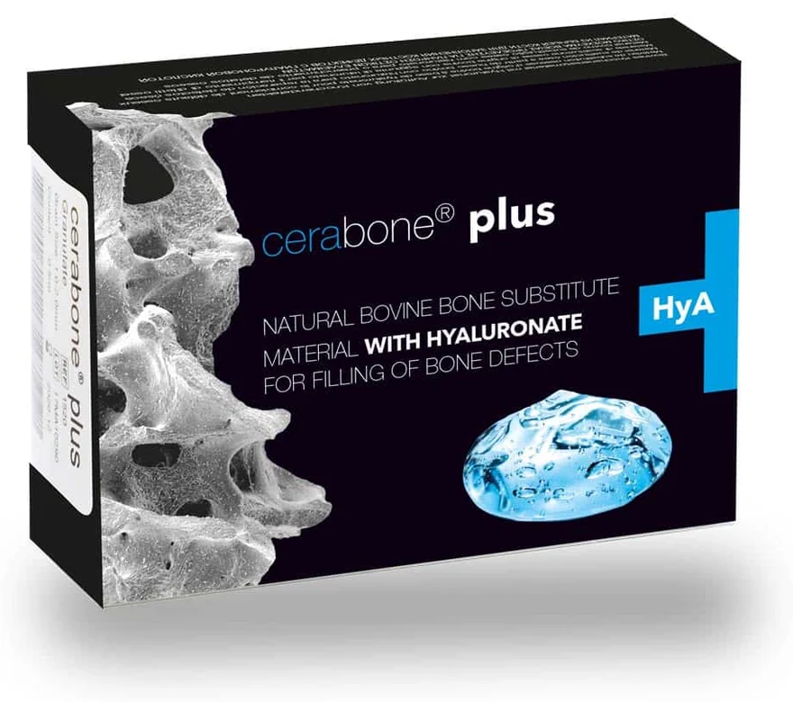
Made of: purified bone mineral from cattle, plus hyaluronic acid made by fermentation. You may also see its earlier name, cerabone® plus.2

The cerabone plus pack, labelled natural bovine bone substitute material with hyaluronate.

The pack, as supplied by the manufacturer.

  • Shaped to fit your jaw

    Ordinary graft granules are loose, a little like wet sand. These hold together, so your dentist can shape the graft precisely and fewer granules drift out of place.1

  • Keeps its shape for years

    The mineral dissolves very slowly. It stays as a lasting framework inside your new bone and helps the area keep its height and width.3

  • Built like your own bone

    Around 65–80% of the material is open space, like a sponge. Blood vessels and bone-forming cells can grow straight into it.3

  • Often no second wound

    Bone does not have to be taken from another part of your mouth. In some cases your dentist may still mix in a little of your own bone to help healing.6

  • Uses a substance your body already makes

    Hyaluronic acid is found naturally in skin, gums and bone. Your body absorbs it within the first weeks.2

The technology, simply

1,200 °C
The bone is heated to this temperature. It removes every organic part and destroys bacteria, viruses and prions, leaving pure mineral.3
1,000 times
Hyaluronic acid can hold roughly this many times its own volume in liquid. That is what binds the granules into one mouldable mass.1
20+ years
The manufacturer reports that cerabone® has been used for more than 20 years and in millions of patients. It has carried the European CE mark since 2002.3
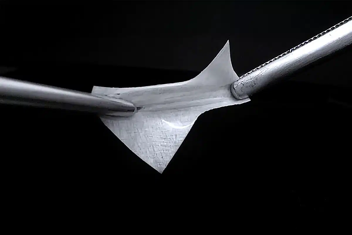
A thin, flexible sheet of Jason membrane held between two surgical instruments.
The membrane itself: a thin, flexible sheet that drapes over the graft.

Jason® membrane

Collagen barrier membrane

A very thin sheet of natural collagen. It is laid over the bone graft to hold the granules in place and to stop gum tissue growing into the space meant for bone.4

Made of: purified collagen from animal pericardium, the strong, flexible tissue that surrounds the heart.5

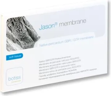
The Jason membrane pack, labelled native pericardium GBR/GTR membrane.

The pack, as supplied by the manufacturer.

  • Thin, so the gum closes without strain

    At about 0.15 mm it is roughly as thin as a sheet of paper. That makes it easier to close the gum over the graft without pulling it tight, which helps most if your gums are naturally thin.4

  • Strong for its size

    Its collagen fibres run in many directions, so the membrane resists tearing. It can be held in place with stitches or tiny pins when needed.4

  • Protects for months

    Its dense, layered collagen breaks down slowly. This gives slow-growing bone more time, which matters most in larger repairs.5

  • Dissolves on its own

    Your body absorbs the membrane. There is no operation to take it out.7

  • No chemical hardening

    Some membranes are chemically treated to make them last longer. Jason® membrane gets its durability from its natural structure instead.4

The technology, simply

~0.15 mm
The typical thickness of the membrane. Because it is a natural tissue, each sheet varies a little.5
100,000 beats
The tissue it comes from flexes with every heartbeat, about 100,000 times a day. That natural toughness is kept during manufacture.5
3–6 months
How long the manufacturer estimates your body takes to absorb it. The estimate is based on laboratory and animal studies.5

When these materials are used

  • After a tooth is removedTo stop the empty socket shrinking, so an implant can be placed later.
  • Before or during implant placementTo widen or raise a jaw ridge that has become too thin or too low.
  • Sinus liftTo add bone height in the upper back jaw, below the sinus.
  • Around an existing implantTo fill a gap where bone does not fully cover the implant.
  • Bone loss from gum diseaseTo help rebuild bone that has been lost around a tooth.

How they compare with other options

No single material is right for everyone. Each option below has its place, and your dentist may combine more than one. Choose an option to see how it differs.

cerabone® +HyA compared with other bone grafts

Your own bone

Bone taken from another part of your mouth or jaw.

Why we may choose cerabone® +HyA
There is no second surgical site, and the amount available is not limited.
Why your own bone may suit you better
It carries your own living bone cells and natural healing signals. Your dentist may recommend it on its own, or mixed with the graft to add this benefit.68

Donor bone

Human bone from a licensed tissue bank.

Why we may choose cerabone® +HyA
Its mineral stays as a lasting framework. That is useful when keeping the shape of the area for the long term matters most.
Why donor bone may suit you better
Your body turns it into your own bone more quickly, so the next treatment step can sometimes happen sooner. It is not of animal origin.6

Synthetic graft

Lab-made minerals similar to the mineral in bone.

Why we may choose cerabone® +HyA
It has the natural structure of real bone and holds its volume for the long term.
Why a synthetic graft may suit you better
It has no animal or human origin. It is gradually absorbed as new bone forms, which suits some cases well.68

Standard cattle-bone granules

The same kind of purified cattle bone mineral, without hyaluronic acid.

Why we may choose cerabone® +HyA
The granules stick together, so the graft is easier to shape and to keep exactly where it is placed.1
What stays the same
Standard granules give the same kind of lasting support. Easier handling is a practical advantage for your dentist. It has not been proven to mean better healing.10

Jason® membrane compared with other membranes

Collagen membranes from skin tissue

Purified collagen from animal skin (dermis).

Why we may choose Jason® membrane
It is thinner and protects for longer. The same manufacturer’s skin-based membrane is about 0.4 mm thick, compared with about 0.15 mm.5
Why a skin-based membrane may suit you better
Your body absorbs it too. It is sometimes preferred for smaller repairs, or where the gum cannot be fully closed.7

Chemically hardened collagen membranes

Animal collagen that is chemically treated (“cross-linked”) to slow its breakdown.

Why we may choose Jason® membrane
Its long protection time comes from its natural structure, with no added chemical treatment. Animal research suggests such treatment may make it harder for tissue and blood vessels to grow into a membrane.4
Why a hardened membrane may suit you better
It is slow to absorb, so protection can last a long time. How well it performs depends on the particular product.

Non-absorbable membranes

A synthetic sheet (PTFE) or a titanium mesh.

Why we may choose Jason® membrane
Your body absorbs it, so no procedure is needed to remove the membrane.7
Why a non-absorbable membrane may suit you better
It holds its shape better, which helps in some large or complex repairs. Some types can be left uncovered while healing, whereas Jason® membrane needs to stay covered by the gum.7

Where the materials come from

You have a right to know exactly what is placed in your body, so here it is plainly.

cerabone® +HyA

Bone mineral from cattle

The bone mineral is of bovine (cattle) origin. Heating it to 1,200 °C removes every organic part, so only the mineral remains. The hyaluronic acid is made by bacterial fermentation and is not taken from animals.23

Jason® membrane

Collagen from animal tissue

The collagen is of animal origin. It comes from the pericardium of veterinary-checked animals raised for the food industry. A multi-step cleaning process removes cells and all non-collagen proteins, and the finished membrane is sterilised.5

Please tell us if this matters to you.If you would prefer to avoid materials of animal origin for religious, ethical or personal reasons, let us know before your treatment. Other options exist, including materials with no animal content, and we will go through them with you.

What the research shows, and what it does not

We want you to have honest expectations. The handling benefits of these materials are well established. Research on healing is encouraging but still at an early stage.

  • Study in patients, 2026

    Firmer implants at four months

    In 16 implants placed straight after a tooth was removed, those with this graft were more stable at four months than those with no graft. The study was small and short, and it could not show how much of the benefit came from the hyaluronic acid.10

  • Laboratory study, 2022

    More new blood vessels

    In a chick-embryo model, bone substitutes with hyaluronic acid grew more new blood vessels than those without. This is encouraging, but it is not proof that patients heal faster.9

  • Animal study, 2021

    A reliable protective barrier

    Over 12 weeks, a pericardium membrane of this type protected the graft as well as an established collagen membrane. It supports the membrane as a sound choice, not as a better one.11

What we cannot promise: faster healing, less discomfort or a higher implant success rate for every patient. Results depend on your health, your mouth and how you care for the area afterwards.

Getting ready for your consultation

The right material starts with the right plan. An examination and X-rays show what needs rebuilding. What you tell us helps us choose.

Please tell us about

  • Any allergies, especially to collagen or hyaluronic acid
  • Your general health and any medicines you take
  • Whether you smoke
  • Whether you would prefer to avoid materials of animal origin

Questions worth asking

What needs rebuilding, which materials suit your case, what your other options are and how long healing will take. These questions are in the list near the end of this page, ready to tick and take to your visit.

Go to my question list

Common questions

Why do I need a bone graft for an implant?

An implant works like the root of a natural tooth. It needs enough bone, in both height and width, to hold it firmly. After a tooth is lost, the bone around it often shrinks. A graft rebuilds the missing volume so the implant can cope with everyday chewing.8

Will it hurt?

The procedure is done under local anaesthetic, so the area is numb while we work. Our center also offers treatment under sedation if you feel anxious.

Afterwards, some tenderness, swelling or bruising is normal and usually settles within a week or two. We will give you aftercare instructions and advise you on pain relief.8

Are these materials from animals?

Yes. cerabone® +HyA contains purified bone mineral from cattle. Jason® membrane is made of purified collagen of animal origin. The hyaluronic acid is made by fermentation, not taken from animals. If animal origin affects your choice, please tell us so we can discuss alternatives.25

Are these materials safe?

Both are CE-certified medical devices. cerabone® is heated to 1,200 °C, which removes all organic material and destroys bacteria, viruses and prions. Jason® membrane goes through a multi-step purification and is sterilised.35

An allergic reaction to either material is possible but extremely rare. Like any surgery, bone grafting also carries general risks such as infection. Your dentist will explain these before treatment.8

Does the graft stay in my jaw for good?

Partly. The hyaluronic acid is absorbed within the first weeks. The mineral granules become surrounded by your own new bone and stay as part of it for the long term, which is what helps the area keep its shape. The membrane is fully absorbed, usually within a few months.25

Does the hyaluronic acid make healing faster?

We cannot promise that. Its proven job is practical: it binds the granules so the graft can be placed more accurately. Laboratory work and one small study in patients suggest it may also support healing, but this has not yet been shown in large patient studies.910

How long does healing take?

It depends on how much bone needs rebuilding. For this type of graft the manufacturer recommends at least six months of healing before the next surgical step. In suitable cases the implant can be placed at the same visit as the graft. Your dentist will give you a timeline for your own case.36

Will I need another operation to remove the membrane?

No. Your body absorbs Jason® membrane, so it does not have to be taken out. You will still have follow-up visits as part of your treatment plan, for example to place the implant if that is done as a later step.7

Can I ask for a different material?

Yes. Tell us your preferences and we will explain which alternatives are suitable for your case. These can include your own bone, donor bone, synthetic grafts and synthetic membranes.

Is there anyone these materials are not suitable for?

They should not be used if you have a known or suspected allergy to collagen or to hyaluronic acid. Your general health, any medicines you take and smoking can all affect how bone heals, so we review your medical history with you before recommending any graft.25

Rebuilding bone for your dental implant

When a tooth has been missing for a while, the jawbone underneath can shrink. An implant needs enough healthy bone to hold it firmly, just as a house needs a solid foundation. Guided bone regeneration helps your body grow that bone back, protected by titanium mesh or titanium-reinforced PTFE.1,2

Tap through the four steps of the diagram to see how your bone is rebuilt, from start to finish.

Watch your bone rebuild

A simplified side view of the jaw

Cross-section of the jaw during guided bone regeneration Four stages. First, a dip in the jawbone where bone has been lost, with a dotted outline of where bone used to be. Second, bone graft granules fill the dip. Third, a curved titanium cover sits over the graft, held with tiny pins, and the gum is closed over it. Fourth, the graft has become new bone, the cover has been removed, and a dental implant with a crown is in place.
  • Gum
  • Your jawbone
  • Bone that was lost
  • Bone graft
  • Protective cover
  • New bone
  • Implant and new tooth
Step 1 of 4

Bone loss

After a tooth is lost, the bone that held it slowly shrinks. The dotted line shows where bone used to be. Now there isn't enough height or width to hold an implant firmly.

Step 2 of 4

Bone graft

Your surgeon gently opens the gum and fills the gap with bone graft material. It works like scaffolding: your body slowly replaces it with your own living bone.

Step 3 of 4

Protective cover

A titanium mesh or titanium-reinforced PTFE cover is shaped over the graft and held still with tiny pins. The gum is closed over the top. The cover keeps the space open and keeps fast-growing gum tissue out.

Step 4 of 4

New bone, then your implant

Over several months your body builds new bone. The cover is then removed, often at the same visit your implant is placed. Your new tooth follows once the implant has healed.

Simplified illustration. Your own treatment plan may differ.

Why would I need new bone?

Losing bone after losing a tooth is very common, and it can be treated. Guided bone regeneration rebuilds missing jawbone in the exact spot where your implant is planned. Your body grows the new bone itself; we give it the right space, scaffold and protection.1

Bone shrinks after tooth loss

Without a tooth root to support it, the jawbone gradually becomes thinner and lower.

Implants need a solid base

Enough bone all around the implant keeps it stable, healthy and natural-looking for the long term.

New bone rebuilds what was lost

It can restore the width, the height, or both, before your implant or at the same time.

Not everyone needs it. If you already have enough healthy bone, your implant can be placed without it. Smaller repairs may only need a soft membrane that dissolves on its own. The materials on this page are used when more support is needed.3 The dissolving membrane we use for smaller repairs is explained in guide 3.

Why does new bone need protection?

Gum cells grow much faster than bone cells. Without protection, soft gum tissue can move into the space before bone has a chance to fill it.

A protective cover, also called a barrier, says "stop" to the gum. It holds the space open and gives slower bone cells the time they need. That is why the treatment is called guided bone regeneration.1

Which cells reach an open space first?
Gum cells Fast: they spread quickly
Bone cells Slow and steady: they need months

With a cover in place, the gum stays on top and the space underneath is kept for bone.

The two materials we use

Both are temporary covers placed over the bone graft. Both hold the space open while your own bone grows, and both are removed once the bone is ready. They just do it in slightly different ways.

Thin, strong and full of small holes

Titanium mesh

The strong scaffold

A very thin sheet of medical-grade titanium, the same metal most dental implants are made of, with a pattern of small holes. Your surgeon shapes it into a sturdy dome that sits over the bone graft while new bone forms underneath.2

Think of it as a custom-fitted dome: it keeps its shape, whatever presses on it from above.

What it does for you

  • Holds its shape

    Titanium is rigid, so pressure from your gum, cheeks or lips can't squash the graft. That makes it a dependable choice when a lot of bone needs rebuilding.1,2

  • Lets nourishment through

    The small holes let blood supply and nutrients from the gum reach the graft, the fuel new bone needs to grow.2

  • Shaped to fit you

    It can be bent and trimmed to recreate the natural shape of your jaw. It can even be designed from your 3D scan and made to measure.2,4

  • Rebuilds height and width

    Research reviews describe it as a predictable way to rebuild bone upwards and outwards at the same time.2,5

  • Forgiving during healing

    Sometimes a small edge shows through the gum while healing. A review of 17 studies found that, even then, the implant could still be placed in almost every case.5

  • Body-friendly

    Titanium is very well tolerated by the body and does not rust or corrode.2

Often paired with a collagen layer. A thin membrane that dissolves on its own is often placed over the mesh for extra protection.4

Smooth membrane with a titanium frame inside

Titanium-reinforced PTFE

The protective shield

A thin, smooth, white membrane made of dense PTFE, a very stable medical material, with a fine titanium frame built inside it. The frame gives it shape; the membrane forms a continuous protective layer over the graft.6

PTFE stands for polytetrafluoroethylene.

Think of it as a tent: the titanium frame is the tent poles, and the PTFE is the weatherproof fabric stretched over them.

What it does for you

  • Shape and cover in one

    The titanium frame holds the shape your new bone needs, while the membrane covers the graft as one continuous layer.7

  • A tight barrier

    Dense PTFE has extremely tiny pores. This helps keep bacteria and gum cells away from the healing graft.6

  • Strong for the whole healing time

    It doesn't dissolve, so its protection doesn't weaken partway through healing. It keeps working until it is removed.6

  • Gentler removal

    Its smooth surface means gum tissue doesn't grow into it, so it usually lifts away easily when it's time to take it out.6

  • Top results for bone height

    A 2022 analysis that pooled 19 studies ranked titanium-reinforced dense PTFE highest of 12 membrane types for regaining lost bone height.8

  • Stable and non-reactive

    Medical-grade PTFE doesn't break down in the body or react with the tissues around it, and it has been used in dentistry for many years.6

Design matters. Some reinforced PTFE meshes have small openings on purpose, and may be combined with a collagen layer for extra cover.9

Side by side

Both do the same essential job: protect the graft and hold the space while new bone grows. Here is how they differ.

Comparison of titanium mesh and titanium-reinforced PTFE
FeatureTitanium meshTitanium-reinforced PTFE
What it isA thin titanium sheet with small holesA smooth dense-PTFE membrane with a titanium frame inside
Holds its shapeYes, it is very rigidYes, thanks to the titanium frame
How it protects the graftA sturdy shield, often topped with a collagen layer that dissolves10A continuous, tight barrier6
Blood supplyHoles let nourishment through from the gumNourishment comes mainly from the bone underneath
FitBent to shape, or made to measure from your 3D scanTrimmed and shaped to your site; made-to-measure versions also exist9
RemovalA short procedure; tissue can grow into the holes, so it takes a little more careA short procedure; it usually lifts away easily
Often chosen forLarger or complex areas; rebuilding height and width togetherRebuilding height or width where a sealed barrier is preferred

What the research says

Study results describe groups of patients. They can't predict your individual result.

Similar results

Two clinical trials comparing the materials head to head found similar bone gain and complication rates.9,10

5 to 6 mm

Average new bone height gained with made-to-measure versions of both, in a 2024 trial.9

About 98%

Of implants placed in the rebuilt bone in that trial bonded successfully, with either material.9

32 trials

A 2023 review of 32 trials found that covers that don't dissolve, like these, gave the best balance of bone gain and complications.11

Which one is right for me?

There is no single best material for everyone. Your surgeon chooses based on the shape and size of the bone loss and how much gum covers it.3

After examining you and your 3D scan, we will recommend the option that suits you, and explain why.

What your surgeon looks at

  • How much bone is missing, and whether height, width or both need rebuilding
  • Where in your mouth the implant will go
  • The thickness and health of your gum
  • Whether a made-to-measure, 3D-designed cover would help
  • Your general health and habits, such as smoking

Your treatment, step by step

  1. Consultation and 3D scan

    We examine your mouth, take a 3D X-ray (CBCT scan) and plan your new bone and implant together.

    First visit
  2. Bone regeneration

    Done under local anaesthetic, so the area is numb. The graft and cover are placed and the gum is closed over them.

    One visit
  3. Healing

    New bone forms over several months. We see you for regular check-ups along the way.

    Usually 4 to 9 months
  4. Cover removal and implant

    The cover is removed, often at the same visit your implant is placed. Your new tooth follows once the implant has healed.

    Once the bone is ready

In some cases the implant can go in at the same visit as the bone graft. We will tell you which plan suits you and give you a personal timeline.3

Helping your new bone heal

  • Avoid smoking. It slows healing and raises the risk of problems.
  • Eat soft foods and chew on the other side for the first weeks.
  • Don't press on the area with your tongue or fingers. If you wear a temporary denture, we will adjust it so it doesn't press on the gum.
  • Follow your cleaning and mouth-rinse instructions carefully.
  • Keep every follow-up appointment, even if everything feels fine.

Call us if you notice

  • Grey or white material showing through the gum
  • Swelling or pain that gets worse after the first few days
  • A bad taste, discharge or a fever

Please don't try to remove anything yourself. Call 079 660 0883.

Words you may hear

Dental terms can sound complicated. Tap a word to see what it means in everyday language.

Guided bone regeneration (GBR)

A treatment that helps your body grow new jawbone in a protected space, usually before or during implant treatment.

Bone graft

Granules placed where bone is missing. They act as scaffolding that your body gradually replaces with its own bone.

Barrier or membrane

A thin cover placed over the bone graft to keep gum tissue out while bone grows.

Titanium mesh

A thin, rigid sheet of titanium with small holes, shaped over the bone graft.

PTFE

Short for polytetrafluoroethylene: a very stable, non-reactive medical material used to make membranes.

Dense PTFE

PTFE with extremely tiny pores that help keep bacteria out. Expanded PTFE has larger pores.

Collagen membrane

A soft membrane that dissolves on its own. Used for smaller repairs, or as an extra layer over titanium mesh.

Biocompatible

Well tolerated by the body, without harmful reactions.

CBCT scan

A 3D X-ray that shows your jawbone in detail, so your treatment can be planned precisely.

Exposure

When part of the cover becomes visible through the gum during healing. Call us if this happens.

Osseointegration

When bone grows tightly onto the implant's surface, locking it firmly in place.

Local anaesthetic

An injection that numbs only the treatment area, so you stay awake but feel no pain.

Questions patients ask

Choose a topic, or open any question to read the answer. Your dental team can explain how each point applies to you.

Does every implant need bone regeneration?

No. It is only needed when there isn't enough healthy bone for the planned implant. Smaller repairs may use a soft membrane that dissolves on its own, while larger repairs need a firmer cover such as titanium mesh or titanium-reinforced PTFE.3

Why do I need a mesh or membrane at all?

When bone graft is placed, the surrounding gum naturally heals faster and tries to fill the space. The cover acts as a barrier: it says “stop” to the fast-growing gum and gives your slower-growing bone cells the time and space to build a solid foundation.1

How long until I get my implant?

Bone needs time to grow and mature, usually several months, depending on how much bone is being rebuilt and how your body heals. In some cases the implant is placed at the same time as the bone graft; in others, once healing is complete.

After the implant is placed, it also needs time to heal before your new tooth is fitted. We will give you a personal timeline at your consultation.

Am I a good candidate for bone regeneration?

Many adults who don't have enough bone for an implant can benefit. Things that can slow healing include smoking, uncontrolled diabetes, untreated gum disease and certain medicines. We will review your health history and 3D scan with you and recommend the safest, most predictable approach.

Can these materials guarantee my implant will succeed?

No material can guarantee a result. These covers help create the right conditions for new bone, but the outcome also depends on careful planning, your general health, how your gum heals and how well the area is looked after.12

Is titanium safe? Could I be allergic to it?

Titanium is one of the most body-friendly metals used in medicine. Most dental implants are made of it, and it has been used for decades in joint replacements and bone plates.2

True titanium allergy is very rare. If you have ever reacted to metals, please tell us so we can discuss it at your consultation.

What exactly is PTFE?

PTFE (polytetrafluoroethylene) is a very stable, non-reactive material. The dense, medical-grade form used for these membranes doesn't break down in the body or react with the surrounding tissues. Membranes like this have been used in dental bone regeneration for many years.6

What do dense, expanded and perforated PTFE mean?

These words describe how open the material is. Dense PTFE has much smaller pores than expanded PTFE, which helps keep bacteria out.6,7 Some reinforced meshes have larger openings made on purpose, and may be combined with a collagen layer.9 Your surgeon chooses the design that suits your repair.

Why not use a membrane that dissolves on its own?

Dissolving (collagen) membranes are a good choice for smaller, well-supported repairs, and we use them when they are the best fit. For larger repairs, especially when bone height needs rebuilding, a soft membrane can sag into the space. Titanium mesh and titanium-reinforced PTFE keep their shape for the whole healing period.3,8

Can I have an MRI scan while the cover is in place?

Titanium and PTFE are not magnetic, and titanium is generally considered safe for MRI scans, although it can cause small image distortions close to the area. Always tell the MRI team about any dental work or implants you have.

Will it hurt?

The procedure is done under local anaesthetic, so the area is numb and you shouldn't feel pain during it. Some swelling and soreness for a few days afterwards is normal and is usually well controlled with the medicine we recommend. Removing the cover later is a much smaller procedure.

Will I feel the mesh or membrane in my mouth?

It sits under your gum, so it isn't visible. Once the gum has healed, most people don't notice it day to day, although the area may feel slightly firm at first. Try not to press on it with your tongue or fingers.

How long does the cover stay in?

Usually about 4 to 9 months, depending on how much bone is being rebuilt. Neither material dissolves, so the cover is removed in a short, planned procedure, often at the same visit your implant is placed.2,6

What if part of it shows through my gum?

This is called exposure, and it is one of the most common issues with any cover used in bone regeneration.8 Please contact us promptly and don't try to remove anything yourself. It can often be managed with gentle cleaning, antiseptic rinses and closer check-ups.

With titanium mesh, a review of 17 studies found that, despite exposure, implants could still be placed in almost all sites.5 With dense PTFE, the tight barrier helps keep bacteria away from the graft.6

What are the main risks?

The most common issue is the gum opening over the cover (exposure). Less often, infection or loss of part of the graft can happen, which may need extra treatment. Techniques that rebuild more bone can also carry more healing complications, so we plan carefully and follow your healing closely.10,11

Contact us promptly if the material becomes visible, pain or swelling gets worse, or you notice discharge or fever.

Scientific references

This page is based on the peer-reviewed studies and clinical guidance below. The small numbers in the text link here. Links open in a new tab.

  1. ReviewRakhmatia YD, Ayukawa Y, Furuhashi A, Koyano K.

    Current barrier membranes: titanium mesh and other membranes for guided bone regeneration in dental applications.

    Journal of Prosthodontic Research. 2013;57(1):3–14. View on PubMed (opens in a new tab)
  2. ReviewXie Y, Li S, Zhang T, Wang C, Cai X.

    Titanium mesh for bone augmentation in oral implantology: current application and progress.

    International Journal of Oral Science. 2020;12(1):37. Read the full article (opens in a new tab)
  3. Clinical guidanceIkumi R, Osugi K, Pitta J.

    Bone defect morphology and appropriate barrier membrane selection.

    ITI Blog (International Team for Implantology). 9 April 2026. Read on the ITI Blog (opens in a new tab)
  4. Clinical trialCucchi A, Vignudelli E, Franceschi D, Randellini E, Lizio G, Fiorino A, Corinaldesi G.

    Vertical and horizontal ridge augmentation using customized CAD/CAM titanium mesh with versus without resorbable membranes: a randomized clinical trial.

    Clinical Oral Implants Research. 2021;32(12):1411–1424. View the article (DOI) (opens in a new tab)
  5. Systematic reviewRasia-dal Polo M, Poli PP, Rancitelli D, Beretta M, Maiorana C.

    Alveolar ridge reconstruction with titanium meshes: a systematic review of the literature.

    Medicina Oral, Patología Oral y Cirugía Bucal. 2014;19(6):e639–e646. Read the full article (opens in a new tab)
  6. ReviewCarbonell JM, Sanz Martín I, Santos A, Pujol A, Sanz-Moliner JD, Nart J.

    High-density polytetrafluoroethylene membranes in guided bone and tissue regeneration procedures: a literature review.

    International Journal of Oral and Maxillofacial Surgery. 2014;43(1):75–84. View the article (DOI) (opens in a new tab)
  7. Clinical trialRonda M, Rebaudi A, Torelli L, Stacchi C.

    Expanded vs. dense polytetrafluoroethylene membranes in vertical ridge augmentation around dental implants: a prospective randomized controlled clinical trial.

    Clinical Oral Implants Research. 2014;25(7):859–866. View the article (DOI) (opens in a new tab)
  8. Analysis of many studiesZhang M, Zhou Z, Yun J, et al.

    Effect of different membranes on vertical bone regeneration: a systematic review and network meta-analysis.

    BioMed Research International. 2022;2022:7742687. Read the full article (opens in a new tab)
  9. Clinical trialCucchi A, Bettini S, Tedeschi L, Urban I, Franceschi D, Fiorino A, Corinaldesi G.

    Complication, vertical bone gain, volumetric changes after vertical ridge augmentation using customized reinforced PTFE mesh or Ti-mesh: a non-inferiority randomized clinical trial.

    Clinical Oral Implants Research. 2024. View the article (DOI) (opens in a new tab)
  10. Clinical trialCucchi A, Vignudelli E, Napolitano A, Marchetti C, Corinaldesi G.

    Evaluation of complication rates and vertical bone gain after guided bone regeneration with non-resorbable membranes versus titanium meshes and resorbable membranes: a randomized clinical trial.

    Clinical Implant Dentistry and Related Research. 2017;19(5):821–832. Read the full article (opens in a new tab)
  11. Analysis of many studiesAlotaibi FF, Rocchietta I, Buti J, D’Aiuto F.

    Comparative evidence of different surgical techniques for the management of vertical alveolar ridge defects in terms of complications and efficacy: a systematic review and network meta-analysis.

    Journal of Clinical Periodontology. 2023;50(11):1487–1519. View on PubMed (opens in a new tab)
  12. Clinical trialCucchi A, Vignudelli E, Fiorino A, Pellegrino G, Corinaldesi G.

    Vertical ridge augmentation (VRA) with Ti-reinforced d-PTFE membranes or Ti meshes and collagen membranes: 1-year results of a randomized clinical trial.

    Clinical Oral Implants Research. 2021. View on PubMed (opens in a new tab)

This page is general information for patients and does not replace a personal consultation. Every patient is different: treatment options, healing times and results vary from person to person. Content reviewed October 2026.

Guided implant surgery

Your implant, planned before it’s placed

A surgical guide is a small, custom-made template that fits over your teeth or gums. It helps your dentist place your dental implant where it was planned on a 3D scan of your mouth.

  • Planned on a 3D scan of your own jaw
  • Made to fit only your mouth
  • Your dentist stays in control throughout

Explore the guide: tap a number

  1. The future tooth

    Planning starts here. Your dentist designs your new tooth first, then places the implant where it can best hold a natural-looking, easy-to-clean tooth.

  2. The surgical guide

    A thin template, usually 3D-printed from medical-grade resin. It is made from your scans, so it fits only your mouth.

  3. Your own teeth

    The guide clips onto your neighboring teeth so it cannot move. If teeth are missing, it can rest on your gums instead. A firm fit is checked before any instrument is used.

  4. The metal sleeve

    A small metal ring sits right over the planned spot. Each instrument passes through it, which controls where the implant goes, its angle and its depth.

  5. The planned implant

    A small post that replaces the tooth root. Its size, position, angle and depth are chosen on your 3D scan before the procedure.

  6. Your jawbone

    The 3D scan shows how much bone you have and where nerves, the sinus and nearby roots are, so the plan keeps a safe distance from them.

Simplified illustration, not to scale. Guide designs vary from patient to patient. The labels in the picture are in English; the numbered list explains each part.

What is a surgical guide?

When a tooth is missing, a dental implant can replace its root. The implant needs to sit in just the right place: in solid bone, a safe distance from nerves and the sinus, and lined up with the new tooth it will hold.

A surgical guide makes that plan easier to follow. It is a thin template, usually 3D-printed, with a small metal sleeve over each planned implant site. During the procedure, the instruments pass through the sleeve. That controls three things at once: where the implant goes, the angle it sits at, and how deep it is placed.1

Think of it like a stencil

Instead of drawing a shape freehand, you trace one that was carefully designed beforehand. The skill of the hand holding the pen still matters, but the result is more predictable.

You may also hear it called guided implant surgery, computer-guided surgery or static computer-aided implant surgery. They all describe the same idea.

Close-up photo inside a patient's mouth. A clear surgical guide is clipped over the lower teeth, with two metal sleeves sitting over a gap where two implants will be placed.
A real surgical guide in place. The clear template clips onto the remaining teeth, and its two metal sleeves sit exactly where two implants were planned.

Your treatment, step by step

Most of the careful work happens before surgery day. On the day itself, your dentist follows a plan that has already been checked.

  1. Step 1 of 6Consultation and 3D scan

    What happens

    Your dentist examines your mouth and takes a cone-beam CT (CBCT) scan, a 3D X-ray of your jaw. It is quick and painless.2 Read the 3D scan guide

    What it means for you

    The scan shows how much bone you have, and where nerves, sinuses and neighboring roots are.

  2. Step 2 of 6Digital impression

    What happens

    A small camera wand, called an intraoral scanner, records the exact shape of your teeth and gums. Sometimes a traditional impression is used instead.

    What it means for you

    This is what lets your guide fit your mouth snugly.

  3. Step 3 of 6Planning on screen

    What happens

    Your dentist combines both scans, designs your future tooth first, then positions the implant to support it, keeping a safe distance from nerves and other structures. Combining scans like this is what international implant experts recommend.3

    What it means for you

    You can see your plan and ask questions before anything is done. The plan also shows whether you need extra bone, called a bone graft, first.4 Read the bone guide

  4. Step 4 of 6Your guide is made

    What happens

    The guide is designed from your plan, usually 3D-printed, and fitted with metal sleeves. Its fit is checked before surgery day.1

    What it means for you

    Your guide is made for your mouth only, and for your plan only.

  5. Step 5 of 6Surgery day

    What happens

    The area is numbed. The guide is put in place and checked for a firm fit, the implant goes in through the sleeve, and the guide is taken out. Read the surgery day guide

    What it means for you

    In suitable cases, the implant goes in through a small opening in the gum instead of a larger cut.5 Sometimes a temporary tooth can be fitted the same day.6

  6. Step 6 of 6Healing and your new tooth

    What happens

    Over the following weeks to months, the implant bonds with your jawbone. Then your final tooth is made and fitted. Read the healing check guide

    What it means for you

    Your care team will give you your personal timeline. Daily cleaning and regular check-ups help your implant last.

What a guide can do for you

A guide turns a carefully checked plan into a more predictable procedure. Open “What the research says” under any point for the details.

  • More precise placement

    Implants placed with a guide usually end up closer to the planned position and angle than implants placed freehand.

    What the research says

    In studies that compared the two methods, guided implants landed significantly closer to the plan than freehand ones.7,8 On average, guided implants end up within about 1 to 1.5 mm of the planned position.9 Fully guided surgery, where every step goes through the guide, is the most accurate kind.10

  • Planned around nerves and sinuses

    Your dentist sees nerves, the sinus and neighboring roots in 3D before surgery, and plans the implant a safe distance away from them.

    What the research says

    International implant experts recommend planning on combined scans and keeping a safety margin around important structures.3 Expert reviews also link guided surgery with fewer problems during the procedure.11 Small differences between plan and result can still happen, so careful checks remain part of every procedure.4,12

  • Your new tooth comes first

    The implant is planned around the tooth it will hold, so your final tooth can look natural, fit your bite and be easy to clean.

    What the research says

    Planning the tooth first and the implant second is a core idea of computer-guided treatment.1 In one small trial followed for five years, guided implants showed slightly less bone loss around them.13

  • Often a gentler procedure

    When your bone allows, the implant can go in through a small opening in the gum instead of a larger cut.

    What the research says

    Several clinical trials found less pain and swelling after this small-opening approach.5,13 But a review of randomized trials judged the overall evidence on comfort to be limited, and not every study found a difference.8,14 Your dentist will choose the safest approach for you.

  • Can mean less time in the chair

    Much of the work is done during planning, so the surgery itself can be shorter.

    What the research says

    In clinical trials, both the small-opening approach and fully guided surgery shortened operating time.5,15 Keep in mind that scans and planning add a step before surgery day.16

  • Fewer surprises

    You can see your plan before treatment and ask questions. In selected cases, a temporary tooth can be ready on surgery day.

    What the research says

    In a recent trial, patients rated fully guided treatment highest for satisfaction.15 In guided-surgery trials, temporary teeth were fitted on the day of surgery when the implants were stable enough.6

The technology, explained simply

Four pieces of digital dentistry work together to turn your scans into a guide that fits only your mouth.

3D dental scan (CBCT)

What it is

A compact scanner that takes a 3D X-ray of your face and jaws.

Why it matters to you

It shows how much bone you have and exactly where nerves and sinuses are, using less radiation than a conventional hospital CT scan.2

Intraoral scanner

What it is

A small handheld camera that makes a precise 3D model of your teeth and gums.

Why it matters to you

It helps the guide fit snugly, and in many cases it replaces impression material.

Planning software

What it is

Software that combines your scans into one 3D model of your mouth.

Why it matters to you

Your dentist decides the implant’s position, angle, depth and size calmly before surgery, rather than during it.4

3D-printed guide

What it is

A custom template printed in medical-grade resin, with metal sleeves that match the implant instruments.

Why it matters to you

It carries the digital plan into your mouth and keeps each step on the planned path.1

Myth or fact?

Test what you know about guided implant surgery. Pick an answer on each card to see what the research says.

With a guide, the implant ends up closer to where it was planned.

Fact

Studies consistently show that guided placement is closer to the plan than freehand placement.8,9

A guide guarantees my implant will last longer.

Myth

Implants placed freehand by experienced dentists also last very well, and studies show similar survival for both methods.16,17 Long-term success depends on your bone and gum health, your general health, and good cleaning and check-ups.

Guided surgery never hurts and never needs stitches.

Myth

The area is numbed, and a small opening may be possible in suitable cases. But less pain and swelling are not guaranteed, and some cases still need an incision and stitches.11,14

The guide stays in my mouth after surgery.

Myth

It is used only during the procedure and removed straight afterwards. It is separate from your implant and from your new tooth.1

My dentist still makes the decisions during surgery.

Fact

The guide is a tool that supports your dentist’s skill and judgment. It does not replace them.4

A guide can replace a bone graft.

Myth

A guide does not create bone. The 3D plan shows how much bone you have, and a graft may still be needed to hold the implant in the right position.4

Is guided surgery right for me?

Guided surgery suits many implant patients, but not every case. A guide is a precision tool, not a replacement for your dentist’s experience.

At your consultation, your dentist will look at

  • How much bone you have, and whether a bone graft is needed first.4
  • The health of your gums and neighboring teeth, and whether the guide can sit firmly.12
  • How wide you can open your mouth. The guide and longer instruments need space, especially for back teeth.18
  • Whether a small gum opening or a regular incision is safer for you, for example when bone grafting is needed.11
  • Your general health, your timeline and your budget. Scans and planning add a step, which may add to the cost.16

Questions to bring to your consultation

Questions about surgical guides are in your question list further down this page, next to the questions from the other guides. Tick the ones you want to ask, then copy them into a note or message.

  • Would a surgical guide help in my case?
  • Will my surgery be fully guided or partially guided?
  • Can I see my 3D plan before treatment?
  • What are my treatment stages, timeline and total cost?

Go to my question list

Common questions

Choose a topic, or open any question to read the answer.

Will the guide feel uncomfortable?

Comfort

The guide is thin and smooth and rests on your teeth or gums. The area is numbed first, so you may feel light pressure while it is put in place, but it should not hurt. It is taken out as soon as the implant is in.

Will it hurt less, and will I recover faster?

Comfort

Often, but not always. In some trials, patients had less pain and swelling when the implant went in through a small opening.5,13 Other studies found little difference.14 Your recovery also depends on your general health, how many implants you have, and whether you need a bone graft.

Is the 3D scan safe?

Safety

A dental CBCT scan uses X-rays, at a lower dose than a conventional medical CT scan.2 Like any X-ray, it should only be taken when the benefit to your treatment outweighs the risk, and only of the area needed.19 Please tell us if you are, or might be, pregnant.

What if the plan and the result are not exactly the same?

Safety

Small differences can happen, usually around a millimeter or so.9 That is why the plan keeps a safety margin around nerves and other structures, and why the guide’s fit is checked before and during the procedure.3

Is guided surgery suitable for everyone?

Suitability

It suits many implant patients. It can be less practical if you cannot open your mouth very wide, especially for back teeth, or if the site needs a large bone graft.11,18 The guide also has to sit securely.12 Your dentist will tell you at your consultation whether it is right for you.

Can a guide be used if I have no teeth left in that jaw?

Suitability

Yes. When teeth are missing, the guide can rest on the gums instead and is held firmly in place during the procedure.1 Your dentist will explain how your guide is supported.

Can I get a new tooth on the same day?

Results

Sometimes. In guided-surgery trials, temporary teeth were fitted on surgery day when the implants were stable enough.6 It is an option only in selected cases.4 Your dentist will confirm on the day whether it is safe for you. If not, a temporary tooth can be fitted later.

What is the difference between fully guided and partially guided surgery?

Results

With partially guided surgery, only the first drill goes through the guide and the rest is done freehand. With fully guided surgery, every drilling step and the implant itself go through the guide. Fully guided placement is the more accurate of the two.10

Can I see my plan before the procedure?

Results

Yes. Because your implant is planned on a 3D model, your dentist can show you the plan on screen and explain where the implant will go and why. It is a good moment to ask any questions you have.

Does guided surgery cost more?

Time and cost

Scans, digital planning and the custom-made guide can add to the cost of treatment.16 Your treatment plan will show the full cost clearly before you decide.

Will my surgery be shorter, and how long does the guide take to make?

Time and cost

The surgery itself can be shorter in some cases,15 but a shorter appointment is not guaranteed, and planning adds time beforehand.12 The guide is designed and made between your scans and your surgery date. Your care team will give you your personal timeline.

Sources

The numbers in the text link to these sources. Tap a number to preview its source without leaving your place. Research articles use more technical language.

  1. Review

    Current state of the art of computer-guided implant surgery (opens in a new tab)

    D'haese J, et al. Periodontology 2000, 2017.
  2. Patient information

    Dental cone beam CT (opens in a new tab)

    RadiologyInfo.org, Radiological Society of North America and American College of Radiology.
  3. Consensus statement

    Accuracy of static computer-aided implant surgery: consensus statements and clinical recommendations (opens in a new tab)

    International Team for Implantology (ITI), 6th ITI Consensus Conference, 2018.
  4. Consensus statement

    Computer-guided implant surgery (opens in a new tab)

    International Team for Implantology (ITI), 5th ITI Consensus Conference, 2013.
  5. Randomized trial

    Flapless versus open flap implant surgery in partially edentulous patients subjected to immediate loading: 1-year results from a split-mouth randomised controlled trial (opens in a new tab)

    Cannizzaro G, Felice P, Leone M, Checchi V, Esposito M. European Journal of Oral Implantology, 2011;4:177-188.
  6. Randomized trial

    Computer-guided versus free-hand placement of immediately loaded dental implants: 1-year post-loading results of a multicentre randomised controlled trial (opens in a new tab)

    Pozzi A, Tallarico M, Marchetti M, Scarfò B, Esposito M. European Journal of Oral Implantology, 2014;7:229-242.
  7. Randomized trial

    The accuracy of single-tooth implants placed using fully digital-guided surgery and freehand implant surgery (opens in a new tab)

    Smitkarn P, et al. Journal of Clinical Periodontology, 2019.
  8. Systematic review

    Static computer-aided, partially guided, and free-handed implant placement: a systematic review and meta-analysis of randomized controlled trials (opens in a new tab)

    Tattan M, Chambrone L, González-Martín O, Avila-Ortiz G. Clinical Oral Implants Research, 2020.
  9. Systematic review

    The accuracy of static computer-aided implant surgery: a systematic review and meta-analysis (opens in a new tab)

    Tahmaseb A, Wu V, Wismeijer D, Coucke W, Evans C. Clinical Oral Implants Research, 2018;29(Suppl 16):416-435.
  10. Systematic review

    Accuracy of implant placement with computer-aided static, dynamic, and robot-assisted surgery: a systematic review and meta-analysis of clinical trials (opens in a new tab)

    Khaohoen A, Powcharoen W, Sornsuwan T, et al. BMC Oral Health, 2024.
  11. Consensus statement

    Group 5 ITI Consensus Report: Digital technologies (opens in a new tab)

    Wismeijer D, Joda T, Flügge T, et al. Clinical Oral Implants Research, 2018;29(Suppl 16):436-442.
  12. Review

    A comprehensive review and update on the current state of computer-assisted rehabilitation in implant dentistry (opens in a new tab)

    Michelinakis G, et al. Primary Dental Journal, 2024.
  13. Randomized trial

    Computer-guided vs freehand placement of immediately loaded dental implants: 5-year postloading results of a randomised controlled trial (opens in a new tab)

    Tallarico M, Esposito M, Xhanari E, Caneva M, Meloni SM. European Journal of Oral Implantology, 2018;11(2):203-213.
  14. Randomized trial

    Comparing patient-reported outcomes and experiences among static, dynamic computer-aided, and conventional freehand dental implant placement: a randomized clinical trial (opens in a new tab)

    Engkawong S, et al. Clinical Implant Dentistry and Related Research, 2021.
  15. Randomized trial

    The efficacy of freehand, pilot drilled and fully guided implant surgery in partially edentulous patients: a randomized control trial (opens in a new tab)

    Alshadidi AAF, Aldosari LIN, Alshehri AHA, et al. PLOS ONE, 2026;21(1):e0341894.
  16. Review of randomized trials

    Clinical applications and effectiveness of guided implant surgery: a critical review based on randomized controlled trials (opens in a new tab)

    Colombo M, Mangano C, Mijiritsky E, Krebs M, Hauschild U, Fortin T. BMC Oral Health, 2017;17:150.
  17. Review and expert white paper

    Beyond accuracy: clinical outcomes of computer assisted implant surgery (opens in a new tab)

    Sadilina S, Vietor K, Doliveux R, et al. Clinical and Experimental Dental Research, 2025.
  18. Systematic review

    Computer technology applications in surgical implant dentistry: a systematic review (opens in a new tab)

    Jung RE, Schneider D, Ganeles J, et al. International Journal of Oral and Maxillofacial Implants, 2009;24(Suppl):92-109.
  19. Patient information

    Dental cone-beam computed tomography (opens in a new tab)

    U.S. Food and Drug Administration.

This page is general information and does not replace a personal consultation. Whether guided implant surgery is right for you depends on your examination and scans. Sources checked 12 October 2026. Last reviewed October 2026.

Implant surgery with the iChiropro, measured turn by turn

At Magic Tooth Dental Center, we place dental implants with the iChiropro from Bien-Air, a Swiss-made surgical motor that your dentist runs from an iPad.1,10 It keeps the drill speed steady, limits the force used on your implant, cools the bone as it works, and saves a record of your procedure.

  • PlanThe steps are matched to your implant.
  • ControlSpeed, force and cooling are set by your dentist.
  • RecordThe details of your procedure are saved to your file.

The technology assists. Your dentist makes the decisions.

Insertion recordIllustration
Illustration of an implant insertion record A line climbs from left to right as the implant is turned into the bone. It does not rise above a dashed upper limit, and it ends at a marked final value.
Try a bone type

The force rises as the implant is turned in. It stays under the limit your dentist has set, and the final value is saved to your record.

  • Force measured as the implant turns
  • Upper limit chosen by your dentist
  • Final value, saved to your record
A simplified drawing of the kind of curve the iChiropro records. It is not a patient's data and not the real app. Left to right is each turn of the implant; height is the twisting force, called torque.

What the iChiropro is

An implant motor is the machine that turns the small drills used to prepare your jawbone, then slowly screws the implant into place. It has to be slow, strong and exact, which makes it very different from the fast drill used for fillings. The iChiropro is the most fully featured motor in Bien-Air's implant range.5

It is the tool, not the implant. The iChiropro is equipment your dentist uses during surgery. The implant that stays in your jaw is a separate device.3,18

Three parts work together

The motor

Bien-Air MX-i LED

A small electric motor with a built-in light. Its electronics keep the speed steady even when the bone pushes back.1

The handpiece

CA 20:1 L Micro-Series

The angled instrument your dentist holds. It gears the motor down so the drill turns slowly and powerfully, at no more than 2,000 turns a minute.8

The app and foot pedal

iChiropro app on an iPad

The app shows each step with its speed, force and cooling settings. During surgery your dentist works by foot pedal, so the screen does not need to be touched.1,7

What it means for you

Each feature starts with what you gain as a patient. Open one to see how the technology does it.

The force is set, measured and limited

Your implant is placed with a measured amount of force that your dentist chooses for your bone. How it worksHide the details

Torque is the twisting force that screws the implant into place. How firmly the implant sits on day one is one of the things your dentist weighs when planning your healing and your new tooth.14 The iChiropro holds its speed steady and delivers the force that was selected, up to 70 Ncm when placing an implant.1,2,5 If the bone pushes back harder, the motor adjusts automatically so the drill does not jam.1

In Bien-Air's own laboratory tests, the force shown on its systems was within about 5% of the force actually delivered, against about 10% for the competitor systems it tested.5

Cooling is built into the instrument

A steady flow of sterile fluid keeps the bone cool while the site is prepared. How it worksHide the details

Drilling creates friction, and bone is sensitive to heat. Research treats about 47 °C as the point where bone starts to be harmed.15 The iChiropro has its own pump, adjustable from 30 to 150 ml of fluid a minute. The fluid runs through sterile, single-use tubing and never touches the pump itself.4,5 In the Bien-Air handpiece the cooling line runs inside the instrument and sprays onto the drill.1,8

A small, well-lit instrument

Easier access to the back of your mouth and a clear, lit view for your dentist. How it worksHide the details

The handpiece has a small, tapered head that leaves more room to see and to work.1 Light is carried to the tip through optical glass, so the working area stays evenly lit at any speed.5,8,9 The instrument is also shorter and lighter than traditional models, which eases the strain on your dentist's hand during a long procedure.1

Hands stay on the surgery, not on the screen

Your dentist moves through each step with a foot pedal, so gloved hands do not need to touch the controls. How it worksHide the details

Speed, direction, cooling and the move to the next step or the next implant are all controlled by foot.1,4,5 Bien-Air notes that this lowers the risk of cross-contamination.1 The motor is built to go through repeated sterilization cycles, and the control unit is supplied with sterile protective film.1,6

Steps matched to your implant and your bone

The drilling steps follow the instructions written for your specific implant, adjusted to how dense your bone is. How it worksHide the details

Every implant brand has its own recommended order of drills, speeds and force. The iChiropro app stores these sequences for many implant systems, shows them one step at a time, and is kept current with free updates.1,7 Your dentist checks the settings and adapts them to you. The bone density can be entered for each implant position and changed at any point in the operation.4,7

Sequences that adapt automatically to bone density are available for supported implant systems.7

Surgery that can follow a 3D plan

When your case is planned on a 3D scan, that plan can be loaded into the motor instead of being typed in again by hand. How it worksHide the details

The app can import operations planned in coDiagnostiX planning software by scanning a QR code, then show the guided-surgery details for each step.1,7 Up to eight implants can be handled in one operation, each with its own settings, which helps when several teeth are replaced in one visit.7

Guided surgery needs a separate treatment plan and a surgical guide, and plan import is only offered for supported implant systems.1,7 The motor does not choose where the implant goes. Your dentist will tell you whether this approach suits your case.

A record of your surgery

What was placed, where, and how firmly can be saved and kept with your dental records. How it worksHide the details

For each implant the app can log the bone density, the insertion force and, where it is measured, a stability reading known as ISQ. The report can be viewed as a graph or a table and exported as a PDF.1,7 For supported implant systems, the implant's barcode can be scanned so that its exact reference is stored with your record.7 That is useful if the implant ever needs attention years later, including at another clinic.

Measured information for the same-day tooth decision

Some implants can carry a temporary tooth the same day; others need to heal first. The recorded readings give your dentist more to go on. How it worksHide the details

Bien-Air has developed a tool for the iChiropro app called ILP, short for Immediate Loading Probability. A machine-learning program analyzes the force curve recorded while the implant was placed and returns a percentage with a color code.1 In a study of 1,320 implants placed by 11 surgeons, the tool agreed with about 90% of the surgeons' “suitable” ratings and about 96% of their “not suitable” ratings.16

ILP is a decision aid, not a diagnosis. Bien-Air describes it as part of a newer app update, so it may not be used in every procedure.1 The decision always rests with your dentist and takes in your bone, your bite and your general health.

What the technology can and cannot do

Good equipment supports good clinical care, but it does not replace it.

It helps your dentist

  • Keep the drill speed and turning force under measured control
  • Keep the bone cool while the site is prepared
  • Follow the steps written for your implant
  • Keep a clear record of what was done

It cannot promise

  • Less pain. Your comfort comes from the anesthetic and your dentist's care.
  • Faster healing. That depends on your health and your procedure.
  • That an implant will succeed. No device can guarantee a result.
  • Better results than every other modern motor. A list of features cannot show that.

How well an implant does depends mainly on your general health, the quality of your bone, careful planning, your dentist's skill and how you look after the implant afterward. Even with appropriate treatment, complications can occur.18

How it differs from a standard implant motor

Every implant motor turns a drill and pumps cooling fluid. The differences are in how much the system guides, adapts and records.

Feature by feature

FeatureA standard implant motoriChiropro
How it is controlledA dial or buttons and a small displayAn iPad screen showing every step, with a foot pedal during surgery
Drilling stepsGeneral programs that the dentist sets by handReady-made sequences for many implant brands, updated free through the app
Adapting to your boneSettings changed by handBone density entered for each implant; the sequence adapts for supported implants
3D surgical planNo import; settings are entered by handImported from compatible coDiagnostiX planning software
Record of the surgeryThe highest force is read from the display and written into the notesAn automatic report with the force curve for every implant, exportable as a PDF
Implant traceabilityRecorded separately by the teamBarcode scanned into the digital record, for supported implants
Same-day tooth decisionThe dentist's judgment, sometimes with a separate measuring deviceThe dentist's judgment, with an added ILP reading where the app version provides it
Accuracy of the force shown, in Bien-Air's laboratory testsAbout ±10% for the competitor systems Bien-Air testedAbout ±5% for Bien-Air systems

The “standard motor” column follows Bien-Air's published comparison of the iChiropro with its own conventional motors,1,5,11 so it describes a typical dial-controlled unit and not any one competitor. Features differ between brands and models. Other modern motors, such as the W&H Implantmed Plus and the NSK Surgic Pro2, also offer controlled speed and force, cooling, lighting and saved treatment data.12,13

Accuracy is worth asking about. Independent laboratory research on other implant motors found that the force actually delivered can fall below the set value after repeated heavy use.17 The iChiropro was not among the motors tested. These are differences in how the equipment works, not proof of better results.

On the day of your surgery

Where the iChiropro fits into your appointment, from planning to the saved report.

  1. Planning

    Before your appointment, your dentist chooses the implant and its position from your X-rays or 3D scan.

  2. Set-up

    The team connects the sterile instruments and cooling line, then loads the settings for your implant and your bone.

  3. Preparing the site

    Once the area is numb, the site is shaped in small steps at preset speeds with constant cooling. You may notice vibration and pressure.

  4. Placing the implant

    The implant is turned in slowly while the motor measures the force at every turn.

  5. The record

    The report is saved to your file, and your dentist explains the next stage of healing.

Words you may hear

Dental teams use a few technical terms. Here is what they mean.

Torque
Twisting force. It describes how much effort it takes to turn the implant into the bone.
Ncm
Newton centimeters, the unit used to measure torque.
Primary stability
How firmly the implant sits in the bone on the day it is placed.
ISQ
A stability score for an implant, recorded when your dentist measures it.
Immediate loading
Fitting a tooth onto an implant on the day it is placed, instead of waiting for it to heal first.
Guided surgery
Surgery that follows a plan made on a 3D scan, using a custom-made surgical guide.
Irrigation
The flow of sterile cooling fluid onto the drill.
Handpiece
The instrument your dentist holds, which carries the drill.

Questions patients ask

Select a question to read the answer.

Is the iChiropro the implant that stays in my mouth?

No. It is the motor system used during surgery. Your dental implant is a separate device placed in the jaw to support a replacement tooth or teeth.3,18

Is an iPad really in charge of my surgery?

No. Your dentist carries out the surgery and operates the instruments. The iPad is the screen used to choose and view the settings. The motor and the cooling pump are run by the iChiropro unit's own electronics, and during surgery your dentist controls them with a foot pedal.4,10 The system is a CE-marked medical device.7

Will my implant procedure hurt?

Implant surgery is usually done under local anesthetic, which numbs the area.19 The motor does not change how numb you are; the anesthetic does that. During the procedure you may notice vibration, gentle pressure and cool water in your mouth.

If you feel anxious about treatment, tell us before your appointment so we can talk through your options.

Does this technology mean less pain or faster healing?

Those benefits cannot be promised from the motor alone. Your dentist will explain how your comfort will be managed and what aftercare you need. Healing depends on your individual health and your procedure.18,19

Does it make my implant more likely to succeed?

It gives your dentist close control over the surgery and a detailed record of it, but no device can promise a result. How well an implant does depends mainly on your general health, the quality of your bone, careful planning, your dentist's skill and how you look after the implant afterward.18

What is torque, and is there a “good” number?

Torque is twisting force, measured in newton centimeters (Ncm). It describes how much effort it takes to turn the implant into the bone. There is no single right number: the target depends on the implant, your bone and your treatment plan. The motor's job is to deliver what your dentist selects and to record what happened.1,7

Why does cooling matter so much?

Bone is living tissue, and an implant only succeeds if that bone stays healthy enough to grow onto it. Friction from drilling produces heat, and research treats about 47 °C as the level where bone begins to be harmed.15 That is why the drill is rinsed with cooling fluid the whole time it is turning.

Can I have a tooth fitted on the same day?

Sometimes, but not automatically. It depends on how firmly the implant sits in your bone when it is placed, where in the mouth it is, and your bite. The force record, and an ILP reading where the app provides one, give your dentist measured information to go on.1,16

Many implants need time to heal before the final tooth is fitted. That is a normal and common part of treatment, not a setback.18

Is guided surgery used for every patient?

No. The iChiropro can work with compatible planning software, but your dentist decides whether this approach suits your case. It needs a separate treatment plan and a surgical guide, which are not part of the motor itself.1 More about guided surgery, in guide 5

What is kept in my surgical record, and is it private?

The report can include each implant's position and reference, the bone density your dentist entered, the force curve, any stability readings taken, and relevant medical notes.7

It is kept with your dental records at Magic Tooth Dental Center and treated as confidential, like the rest of your clinical notes. Ask your dentist if you would like to see it.

Does it work with the implant brand chosen for me?

The app's library covers many of the leading implant brands, and your dentist can program a custom sequence for any other system.1,7 A few automated features, such as barcode scanning and plan import, are only offered for supported brands.7

How is the equipment kept sterile?

The motor is designed to withstand repeated sterilization cycles.1 The cooling fluid travels through its own sterile, single-use line and never touches the pump.4,5 The control unit comes with sterile protective film, and it is operated by foot during surgery.1,6

Are other modern implant motors less safe?

A list of features cannot show that. Other modern systems also provide controlled placement and digital records.12,13 The iChiropro offers a particular combination of iPad controls and planning tools. Equipment does not replace clinical skill.

What helps my implant last?

Your general and oral health, whether you smoke, how well you clean around the implant and regular dental check-ups all matter. Follow your dentist's personal advice. Even with appropriate treatment, complications can occur.18

References and further reading

Equipment features come from the manufacturer's published information. The patient benefits above are plain-language explanations of those features, not findings from a clinical trial comparing motors. Links open in a new tab.

About the iChiropro

  1. Bien-Air Dental. iCHIROPRO: your ultimate implantology system. Manufacturer product page.
  2. Bien-Air Dental. iCHIROPRO implant set with CA 20:1 L Micro-Series. Features and technical data.
  3. Bien-Air USA. iChiropro. iPad interface, components and digital workflow.
  4. Bien-Air Dental. iChiropro instructions for use (PDF). REF 2100291-0011, March 2025. Controls, irrigation, hygiene and report export.
  5. Bien-Air Dental. CHIROPRO PLUS. Technology notes on torque accuracy, irrigation and foot pedal, and the comparison table for Chiropro, Chiropro Plus and iChiropro.
  6. Bien-Air Dental. iCHIROPRO set with wireless foot pedal. Set contents.
  7. Apple App Store. iChiropro 2 app by Bien-Air Dental SA. App description and version history.
  8. DD Group. Bien-Air CA 20:1 L Micro-Series contra-angle. Distributor product overview and technical data.
  9. BioHorizons. CA 20:1 L Micro-Series contra-angle with LED. Distributor product description.
  10. Dentistry.co.uk. A revolution in implant motor design. 1 April 2014.

Other implant motors

  1. Bien-Air USA. ProImplant and system comparison. Published differences between the console and iPad systems.
  2. W&H. Implantmed Plus and Classic. Control functions, illumination and documentation.
  3. NSK. Surgic Pro2. Torque control, irrigation, LED and iPad data connectivity.

Research

  1. Degidi M, Daprile G, Piattelli A, Iezzi G. Development of a new implant primary stability parameter: insertion torque revisited. Clin Implant Dent Relat Res. 2013;15(5):637–644.
  2. Raj R, Manju V, Kumar-Gopal V, Eswar M. Analysis of factors determining thermal changes at osteotomy site in dental implant placement: an in-vitro study. J Clin Exp Dent. 2021;13(3):e234–e239.
  3. Degidi M, Daprile G, Battelli F, et al. The use of an artificial intelligence-driven novel tool for the evaluation of dental implants primary stability and immediate loading feasibility: a multicenter retrospective study. J Clin Med. 2025;14(6):2011. doi:10.3390/jcm14062011.
  4. Lee DH, Cho SA, Lee CH, Lee KB. The overuse of the implant motor: effect on the output torque in overloading condition. Clin Implant Dent Relat Res. 2015;17(3):435–441. doi:10.1111/cid.12195.

General information about dental implants

  1. U.S. Food and Drug Administration. Dental implants: what you should know. Healing, ongoing care, benefits and risks.
  2. Cambridge University Hospitals. Insertion of dental implants. Patient information about the procedure and local anesthesia.

This page gives general information for patients of Magic Tooth Dental Center and does not replace a personal consultation. The features in use depend on the installed equipment, the software version and the implant system chosen for you.

Product and brand names belong to their respective owners. Sources checked .

Implant stability check with Osstell Beacon

Is your implant ready for its new tooth?

A dentist holds the Osstell Beacon reader just above a small peg fitted to an implant in a patient’s lower jaw. A green light glows near the tip of the reader.
Osstell Beacon in use. The reader is held close to the implant and shows a green light for a higher reading.

After an implant is placed, your jawbone needs time to grow around it and hold it firmly. At Magic Tooth Dental Center we use a small handheld device called Osstell Beacon to check how firmly it is held. The check takes seconds and gives a number that helps your dentist choose the right time for the next step.1

  • Over in secondsEach reading takes a second or less once the small peg is in place.4
  • No X-raysThe reading uses gentle magnetic pulses, so it adds no radiation.3
  • A number you can followFrom 1 to 99. Your dentist can explain it and compare it between visits.2

Why timing matters

A dental implant is a small post that sits in the jawbone and does the job of a tooth root. Over the following weeks and months, bone grows onto it and holds it in place. Your new tooth, the crown, is fitted once the implant is held firmly enough for chewing.8

Too soon

Chewing on an implant before the bone has a firm hold can disturb healing.3

Timed to your healing

A stability reading shows how your own implant is doing, so the timing can follow your healing and not a fixed calendar.1

Longer than needed

Waiting a standard “just in case” period can mean more time without your tooth than your healing calls for.1

Everyone heals at a different pace. It depends on things like your bone, where in the mouth the implant sits, and your general health.10

What happens during the check

The check is done in the dental chair as part of a normal implant visit. There are three short steps.

How the stability check works A dental implant sits in the jawbone between two teeth. A small peg is attached to the top of the implant. A handheld reader is held close to the peg without touching it and shows a reading of 72 with a green light. Reading 72 Peg Reader Implant Jawbone
The reader is held close to a small peg on the implant and never touches it. Simplified drawing, not to scale.
  1. A small peg is fitted

    Your dentist gently screws a small sterile peg, called a SmartPeg, onto the implant using light finger pressure. It has a tiny magnet on top.4

  2. The reader is held close

    The handheld reader is held a few millimetres from the peg and does not touch it. It sends gentle magnetic pulses that make the peg vibrate very slightly.6

  3. A number appears

    The reader turns that vibration into a number and shows a red, yellow or green light. Your dentist takes a reading from two directions, then removes the peg.14

Think of a guitar string. A tight string vibrates quickly and gives a high note. A slack string vibrates slowly. In the same way, a peg on a firmly held implant vibrates faster, and the reader shows a higher number.3

Dentists call this method resonance frequency analysis. It was first described in the 1990s and has been studied widely since.78

Do you have a pacemaker or another implanted electronic device? Please tell us before your visit. The reader uses a magnetic field, so your dentist needs to know before doing the check.6

What your number means

The number is called an ISQ, short for Implant Stability Quotient. It runs from 1 to 99, and a higher number means the implant is held more firmly. Dentists commonly read it in three bands.2

  • Lower stability Below 60, red light

    The implant usually needs more healing time before a tooth is fitted. This is common early on and does not, by itself, mean something is wrong. Your dentist will plan another check.

  • Medium stability 60 to 69, yellow light

    The implant is held, but not yet as firmly as it could be. Your dentist may suggest a little more healing time, or a careful step-by-step approach.

  • Higher stability 70 and above, green light

    The implant is firmly held at this visit. Your dentist looks at this together with the rest of your examination before moving to the next step.

  • It is not a percentage

    A reading of 70 does not mean “70% healed”. It is a point on a firmness scale.3

  • Two directions, and the lower one counts

    Bone support can differ around an implant, so it is measured from two directions. Your dentist goes by the lower number.4

  • Most readings are between 55 and 80

    Implants in the lower jaw often read a little higher than those in the upper jaw.2

  • A guide, not a guarantee

    A high number is reassuring, but research has not shown that the first number on its own can predict how an implant will do in the long term. Your gums, bone, bite and general health all matter too.13

Following your healing over time

One reading is a snapshot. Comparing readings from different visits shows which way your healing is heading.5

How implant stability often changes during healing A line starts at a medium level on placement day, dips slightly during early healing, then rises through later healing and levels off before the new tooth is fitted. FirmerTime 1 2 3 4
Illustration only. Real readings and healing times differ from person to person.
  1. Placement day

    A first reading gives a starting point to compare with later.5

  2. Early healing

    As the bone around the implant renews itself, the reading can dip a little before it climbs. A small dip usually levels out.28

  3. Later healing

    New bone takes a firmer hold and the reading usually rises. If it drops a lot instead, your dentist will look into why and may allow more time.2

  4. Before your new tooth

    A reading before the final tooth is fitted helps your dentist confirm the timing.5

How it compares with other checks

Dentists have several ways to judge an implant. Each one answers a different question, and they are often used together. Choose a check to see how it differs.

Feel and tapping

What it tells your dentist
A trained impression of how solid the implant feels, or sounds when tapped with an instrument.
Its limits
It depends on personal judgement and cannot be written down as a number.9
What Osstell Beacon adds
A measured number alongside your dentist’s judgement, which makes changes between visits easier to see.1

Insertion torque

What it tells your dentist
How much resistance the implant met as it was screwed into the bone.
Its limits
It can only be measured once, on the day the implant is placed.12
What Osstell Beacon adds
A reading that can be repeated later without turning the implant. The two measures are related but not the same, so they work well together.13

Dental X-rays

What it tells your dentist
A picture of the bone around the implant and of the implant’s position.
Its limits
X-rays show shape and bone level, not how firmly the implant is held.9
What Osstell Beacon adds
A firmness reading with no X-ray exposure. X-rays remain an important part of implant care and are still used when they are needed.3

Tapping devices, such as Periotest

What it tells your dentist
A number based on how the implant responds to a series of light, rapid taps.
Its limits
It works by tapping the implant, or a part fixed to it, and its numbers use a different scale.11
What Osstell Beacon adds
A reading taken without contact. In a three-year study the two methods agreed only partly, so their numbers cannot be swapped. Both can be useful.11

Reverse torque test

What it tells your dentist
Whether the implant resists a set twisting force. The answer is pass or fail.
Its limits
The twisting force can itself disturb an implant that is still healing, so it is not suited to routine checks.9
What Osstell Beacon adds
A reading that puts no twisting force on the implant and can be repeated while it heals.3

Other vibration-based devices

What it tells your dentist
A stability number from the same vibration principle, measured with a different brand of reader.
Its limits
Readings from different brands can differ by a few points, so it is best to follow your healing with the same system.12
How Osstell Beacon differs
The ISQ scale comes from Osstell and has been used in more than 1,500 published studies. A laboratory study in animal jawbone found the brands broadly agreed, with some differences in how repeatable the readings were. It did not test results in patients, so it cannot say that any one brand is better for patients.112

Questions patients often ask

Does the check hurt?

The reading itself is taken without contact. The reader never touches your implant, and nothing is drilled or injected for it. Fitting the small peg involves light finger pressure. If the area is tender, tell your dentist so they can take extra care.34

How long does it take?

Each reading takes a second or less. Readings are taken from two directions, so once the peg is in place the measuring is over in moments.4

Is it safe? Does it use X-rays?

The check uses a low-energy magnetic field, not X-rays, so it adds no radiation.3

Because the field is magnetic, please tell us beforehand if you have a pacemaker or any other implanted electronic device.6

Does a high number mean my implant will definitely succeed?

A high number is a good sign for stability on that day, but it cannot promise long-term success. A 2026 review of 48 studies did not find that the first number, on its own, predicts how an implant will do later. Healthy gums, good cleaning, your bite and regular check-ups all matter too.13

Does a low number mean my implant has failed?

No. One lower reading does not diagnose failure. Early in healing it often just means the bone needs more time. Your dentist may repeat the reading at a later visit, allow longer healing, or look more closely if the number keeps falling.2

Why were two numbers taken?

Bone support is rarely the same all the way around an implant. Measuring from two directions shows the firmer and the less firm side, and your dentist goes by the lower number.4

When will my implant be measured?

A reading is often taken on the day the implant is placed, as a starting point, and again before the final tooth is fitted. Your dentist will decide whether extra readings in between would help.5

Can it help me get my new tooth sooner?

Sometimes. If the readings show your implant is firmly held, your dentist may be able to go ahead without waiting a fixed standard period. The check does not speed up healing, and an earlier tooth is not right for everyone.113

Can every implant be measured, even after the crown is on?

The peg has to match your type of implant and be able to reach it. If a healing cap or crown is in the way, it may need to come off first. Some implants can be measured through the connector piece instead, although those numbers are read a little differently. Your dentist will tell you what is possible in your case.411

Does it replace X-rays or my check-up?

No. It adds one piece of information. Your dentist still relies on your examination, and on X-rays when they are needed, to see the full picture.9

Is there an extra cost?

Please ask our team how stability checks fit into your implant treatment plan. We will explain each step and its cost before you start.

Sources

The small numbers in the text link to the sources below. How the device works is taken from the manufacturer’s own information. Comparisons and limits are taken from published research. Links open in a new tab. Checked on .

From the manufacturer

  1. Osstell Beacon: product overviewOsstell AB. What the device is for and how it is used.
  2. The Osstell ISQ scaleOsstell AB. The 1 to 99 scale, the three bands and the usual range of readings.
  3. The technique behind OsstellOsstell AB. How the magnetic pulses and vibration measurement work.
  4. SmartPeg support and reference guideOsstell AB. How the peg is fitted, and why two directions are measured.
  5. Osstell clinical guidelinesOsstell AB. When readings are usually taken during treatment.
  6. Osstell Beacon: instructions for use (PDF)Osstell AB. Warnings and precautions, including pacemakers.

Published research

  1. Meredith N, Alleyne D, Cawley P. Quantitative determination of the stability of the implant–tissue interface using resonance frequency analysisClinical Oral Implants Research. 1996;7(3):261–267. The study that introduced the method.
  2. Sennerby L, Meredith N. Implant stability measurements using resonance frequency analysisPeriodontology 2000. 2008;47:51–66. A review of how stability changes as an implant heals.
  3. Atsumi M, Park SH, Wang HL. Methods used to assess implant stability: current statusInternational Journal of Oral & Maxillofacial Implants. 2007;22(5):743–754. A review of the different checks.
  4. Huang H, Wu G, Hunziker E. The clinical significance of implant stability quotient (ISQ) measurements: a literature reviewJournal of Oral Biology and Craniofacial Research. 2020;10(4):629–638. What affects the readings.
  5. Reynolds I, Winning L, Polyzois I. A three-year prospective cohort study evaluating implant stability utilising the Osstell and Periotest devicesFrontiers in Dental Medicine. 2023;4:1139407. A study in 29 patients. It used an earlier Osstell reader, not the Beacon model.
  6. Blazquez-Hinarejos M, et al. Reliability and agreement of three devices for measuring implant stability quotient in the animal ex vivo modelApplied Sciences. 2021;11(8):3453. A laboratory comparison of Osstell Beacon, Penguin and MegaISQ in animal jawbone, not in patients.
  7. Tisci A, et al. Resonance frequency analysis and clinical outcomes in implant dentistry: a systematic review and meta-analysisClinical Implant Dentistry and Related Research. 2026;28(3):e70156. A review of 48 studies on what the readings can and cannot predict.

This page gives general information for patients and does not replace advice from your dentist. Treatment decisions are made for each person individually. Osstell, Osstell Beacon, ISQ and SmartPeg are trademarks of Osstell AB. Periotest, Penguin and MegaISQ are trademarks of their owners. Magic Tooth Dental Center is not affiliated with these manufacturers.

Back to the top

A gentler way to make your implant crown

Once your implant has healed, the next step is the crown: the tooth-coloured part you will see and chew with. To make it, we record your mouth with the 3Shape TRIOS® scanner instead of a tray of impression paste. A slim wand takes thousands of pictures and turns them into a 3D model of your teeth and gums, right on the screen.

  • No impression paste
  • Breathe and swallow normally
  • Pause whenever you need
  • Uses light, not X-rays
Illustration of a digital scan building a 3D model of the upper teeth, with the implant site marked Your implant

Simplified illustration of a scan building up on screen.

Your new tooth has three parts

An implant tooth is built in layers. Choose a part to see what it does and why we scan around it.

Ceramic crown

The tooth-coloured top that you see, smile with and chew with. It is shaped and shaded to match your neighbouring teeth.

Why we scan: the laboratory needs the exact space your crown will fill and how your upper and lower teeth meet.

Abutment

A small connector that joins the crown to the implant and rises through your gum.

Why we scan: the scan records the shape of your healed gum, so the abutment and crown come out of it naturally.

Implant

A small post, usually titanium, that sits in your jaw bone in place of the tooth root. It is hidden under the gum.

Why we scan: to show the scanner exactly where your implant is, we attach a small marker called a scan body for a few minutes.

What your scan records

  • The position and angle of your implant
  • The shape of your gum around it
  • Your neighbouring teeth
  • How your upper and lower teeth bite together
Diagram of an implant tooth showing, from top to bottom, the ceramic crown, the abutment, the gum, the implant and the jaw bone Ceramic crown Abutment Gum Implant Jaw bone (connector) (new tooth root)
The crown is shown lifted so you can see the abutment underneath.
The traditional way

Tray and impression paste

A tray of soft paste is pressed over your teeth. You hold still for a few minutes while it sets, then it is removed and sent to the laboratory as a physical mould.

The TRIOS way

A digital scan

A slim scanner tip glides over your teeth and gums. The pictures build a live 3D model on screen, and the finished file is sent to the laboratory digitally.

Meet the 3Shape TRIOS® scanner

TRIOS is made by 3Shape, a Danish digital-dentistry company.1 Here is what it does, in plain language.

It takes thousands of pictures

The wand shines light onto your teeth and captures images very quickly. Software joins them together into an accurate 3D model as we go.1

It sees in real colour

Your teeth and gums appear in natural colour, and the scanner can record tooth shades to help match your new crown.4

It is made for implants

A small scan body attached to your implant shows the scanner the implant's exact position and angle beneath the gum.

It checks as it goes

Your dentist watches the model build on screen. If a small area is missed, only that spot is rescanned, with no need to start again.

It sends your scan digitally

The finished scan reaches the dental laboratory as a secure digital file, so work on your crown can begin straight away.

It is hygienic by design

The part that goes in your mouth is either a single-use tip or a tip that is heat-sterilised between patients.2,3

What happens at your scan visit

Most people find it quick and easy. Step through the visit below to see what we do and what you will feel.

Step 1First visit

We check your implant and prepare the area

Your dentist checks that the gum around your implant has healed well, then gently removes the healing cap (the small cap that has been shaping your gum).

What you will feel: a moment of light pressure. The scan itself does not usually need an injection.

Step 2First visit

We scan the shape of your gum

The scanner tip glides over your teeth and the gum around the implant. This shape guides how your new crown will come out of the gum, just like a natural tooth.

What you will feel: a smooth tip moving over your teeth. You can breathe through your nose and swallow normally.

Step 3First visit

We find the exact position of your implant

A small scan body is attached to the implant for a few minutes and scanned. It tells the software exactly where your implant sits and at what angle. Then it comes off and your healing cap goes back on.

What you will feel: light pressure for a moment while it is attached. If your abutment is already fitted, we may scan it directly instead.

Step 4First visit

We record how your teeth meet

We scan your opposite jaw, then you close your teeth together gently while we scan from the side. This records your bite.

What you will feel: nothing more than biting normally and holding still for a few seconds.

Step 5Second visit

Your crown is made, then fitted

Your scan goes to the dental laboratory, where your crown is designed on computer and made. At your next visit we fit it and check the fit, bite, colour and comfort.

Good news: in a clinical study, implant crowns made from digital scans needed less adjusting on fitting day.10

Step 1 of 5

Why patients prefer the scan

Digital implant impressions have been compared with traditional ones in clinical studies. Here is what they found.

Digital scan and traditional impression, side by side

  • Digital scan
  • Traditional impression

How convenient patients rated it (score out of 100, higher is better)5

Digital79
Traditional54

Time to take the implant impression (minutes, lower is better)5

Digital15 min
Traditional18 min

Time spent adjusting the crown on fitting day (minutes, lower is better)10

Digital2.2 min
Traditional6 min
20 of 20

patients in a randomised implant trial said they would choose the digital scan next time, after trying both methods.5

The time bars use a 0–20 minute scale (impression) and a 0–8 minute scale (adjusting). These studies compared digital scanning in general with traditional impressions, not one scanner brand, and involved small groups of patients. Your own experience may differ.

Showing all 10 benefits
Comfort

Easier if you gag easily

Nothing fills the back of your mouth. A review of patient studies found less nausea, unpleasant taste and breathing difficulty with scanning than with impression paste.7

Comfort

No unpleasant taste

There is no impression material, so there is nothing to taste. In a trial of 40 implant-crown patients, the digital group reported less taste irritation.6

Comfort

See and understand

Your 3D scan appears on screen, so we can show you your implant site, gums and bite, and answer your questions as we go.

Time

A shorter impression

Digital implant impressions took about 15 minutes against 18 in one trial, and about 11 against 14½ in another.5,8

Time

Less work behind the scenes

In one study, clinic and laboratory time together came to about 185 minutes for digitally made implant crowns, against 223 minutes for traditional ones.9

Time

Less adjusting at fitting

Implant crowns made from digital scans needed about 2 minutes of adjusting on fitting day, compared with 6 minutes for traditionally made crowns.10

Results

Accurate for single crowns

Research reviews find digital scans accurate enough for single implant crowns and small bridges in everyday practice.11,13

Results

A natural-looking match

Colour detail of your gums and neighbouring teeth helps the technician design a crown that blends in.4 We always confirm the shade with you in person.

Safety

Light, not X-rays

The scanner records the surface of your teeth with light, much like a camera. It does not use X-rays.1

Safety

A clean tip every time

Scanner tips are either single-use or heat-sterilised between patients.2,3

Traditional impression or TRIOS scan?

The two methods at a glance, from your side of the chair.

Comparison of a traditional impression and a TRIOS digital scan
What it is likeTraditional impressionTRIOS digital scan
What goes in your mouthA tray filled with impression pasteA slim scanner tip
TasteThe paste can taste unpleasantNothing to taste
Gag reflexPaste can spread towards the back of the mouthEasier to tolerate, and we can pause any time
Holding stillA few minutes while the paste setsScanned in sections, with breaks if needed
If something is missedThe impression is usually taken againOnly the missed spot is rescanned
Getting to the laboratoryA physical mould is packed and deliveredA secure digital file
On the wayA mould can distort or breakA file cannot shrink or crack
Seeing the resultNot until the crown is madeA live 3D model on screen

Myth or fact?

Test what you know about digital scans. Pick an answer to see the explanation.

“The scanner uses X-rays.”

It is a myth.TRIOS records the surface of your teeth with light. Separate dental X-rays may still be needed to check the implant and the bone around it.

“You can breathe and swallow normally during the scan.”

It is a fact.Nothing blocks the back of your mouth, and we pause whenever you raise your hand.

“If one spot is missed, the whole scan has to be redone.”

It is a myth.Your dentist sees any gap on screen straight away and rescans only that area.

“A digital scan means I get my final crown the same day.”

It is a myth.Your crown is made by a dental laboratory, so the crown stage usually takes two visits.9,10

Frequently asked questions

Search for a word, or open any question below.

Does the scan hurt?

No. The scanner simply glides over your teeth and gums; it does not press or pinch. Taking off the healing cap and attaching the scan body may feel like light pressure for a moment. If any area feels sensitive, tell us and we will pause.

Is it safe? Does the scanner use X-rays?

The scanner works with light, much like a camera, and does not use X-rays. The tip that goes in your mouth is either single-use or heat-sterilised between patients.1,2,3 You may still need separate dental X-rays so we can check your implant and the bone around it.

How long does the scan take?

The scanning itself usually takes only a few minutes. In clinical studies of single implant crowns, the whole digital impression procedure took about 11 to 15 minutes on average.5,8 Your appointment may be longer if other checks are needed.

I gag easily. Can I still have a digital impression?

Yes, and many people with a strong gag reflex find it much easier. Nothing fills the back of your mouth, you can breathe through your nose and swallow, and we stop the moment you raise your hand.7

Will I still need impression putty?

Usually not. For most single implant crowns, a digital scan replaces the traditional impression. For more extensive work, such as several implants across a whole jaw, your dentist may recommend a traditional or combined approach.

Is a digital scan as accurate as a traditional impression?

For single implant crowns and small bridges, research reviews consider digital scans accurate and reliable in everyday practice.11,13 Some laboratory studies measured traditional impressions as slightly more precise in certain situations, but digital scans were still judged suitable.12 Accuracy also depends on scanning technique, which is one reason your dentist checks the model on screen as they go.13

What is a scan body?

A small, precisely shaped marker that is attached to your implant for a few minutes. The software knows its exact shape, so scanning it shows the position and angle of your implant beneath the gum. It comes off straight after the scan.

How many visits will I need? Can I get my crown the same day?

The crown stage usually takes two visits: one for the scan and one for fitting. Studies of single implant crowns found the same with digital and traditional methods.9,10 The digital difference is less time in the chair and, typically, less adjusting on fitting day.10

Will my crown look natural?

The scan captures your teeth and gums in colour, which helps the laboratory design a crown that matches.4 The final look also depends on the ceramic, shade matching and the technician's work, so we also check the shade with you in person.

Can I see my scan?

Of course. Your 3D model appears on screen during the scan, and we are happy to show you your implant site, your gums and your bite.

What happens to my scan data?

Your scan is stored securely as part of your dental record and shared only with the dental laboratory making your crown.

Can my scan be reused if my crown ever needs replacing?

Your digital record stays on file and can help if a crown ever needs to be remade. If your mouth has changed since, we may take a quick new scan to make sure the fit is right.

Sources

The numbers next to each claim link here. Clinical studies compare digital intraoral scanning in general with traditional impressions; manufacturer sources describe product features as stated by 3Shape.

About the scanner

  1. 3Shape A/S. TRIOS intraoral scanners [manufacturer web page]. 3shape.com/en/scanners/trios
  2. 3Shape A/S. TRIOS accessories: single-use Ready Tips, autoclavable scanner tips and single-use sleeves [manufacturer web page]. 3shape.com/en-gb/scanners/accessories
  3. 3Shape A/S. 3Shape launches all-new TRIOS 5 Wireless intraoral scanner [press release]. October 2022. news.cision.com
  4. Dental Products Report. 3Shape releases TRIOS 3 digital impression solution (colour scanning and shade recording) [news report]. dentalproductsreport.com

Patient experience

  1. Joda T, Brägger U. Patient-centered outcomes comparing digital and conventional implant impression procedures: a randomized crossover trial. Clin Oral Implants Res. 2016;27(12):e185–e189. doi:10.1111/clr.12600
  2. Kunavisarut C, Jarangkul W, Pornprasertsuk-Damrongsri S, Joda T. Patient-reported outcome measures (PROMs) comparing digital and conventional workflows for treatment with posterior single-unit implant restorations: a randomized controlled trial. J Dent. 2022;117:103875. doi:10.1016/j.jdent.2021.103875
  3. The Dental Elf. Intraoral scanning v conventional impressions: patient related outcomes [evidence summary of a systematic review and meta-analysis]. nationalelfservice.net
  4. Guo DN, Liu YS, Pan SX, et al. Clinical efficiency and patient preference of immediate digital impression after implant placement for single implant-supported crown. Chin J Dent Res. 2019;22(1):21–28. doi:10.3290/j.cjdr.a41771

Time and fit

  1. Joda T, Brägger U. Time-efficiency analysis comparing digital and conventional workflows for implant crowns: a prospective clinical crossover trial. Int J Oral Maxillofac Implants. 2015;30(5):1047–1053. doi:10.11607/jomi.3963
  2. Joda T, Katsoulis J, Brägger U. Clinical fitting and adjustment time for implant-supported crowns comparing digital and conventional workflows. Clin Implant Dent Relat Res. 2016;18(5):946–954. doi:10.1111/cid.12377

Accuracy

  1. Afrashtehfar KI, Alnakeb NA, Assery MKM. Accuracy of intraoral scanners versus traditional impressions: a rapid umbrella review. J Evid Based Dent Pract. 2022;22(3):101719. doi:10.1016/j.jebdp.2022.101719
  2. Albanchez-González MI, Brinkmann JC-B, Peláez-Rico J, López-Suárez C, Rodríguez-Alonso V, Suárez-García MJ. Accuracy of digital dental implants impression taking with intraoral scanners compared with conventional impression techniques: a systematic review of in vitro studies. Int J Environ Res Public Health. 2022;19(4):2026. doi:10.3390/ijerph19042026
  3. Ma J, Zhang B, Song H, Wu D, Song T. Accuracy of digital implant impressions obtained using intraoral scanners: a systematic review and meta-analysis of in vivo studies. Int J Implant Dent. 2023;9:48. doi:10.1186/s40729-023-00517-8

This page is general patient information and does not replace a personal consultation. Your dentist will recommend the impression method that suits your mouth and your treatment.

3Shape® and TRIOS® are registered trademarks of 3Shape A/S, named here to identify the equipment used at Magic Tooth Dental Center. Magic Tooth Dental Center is not affiliated with or endorsed by 3Shape. Illustrations are simplified and do not show a specific product. Content reviewed October 2026. Back to top

Questions to take to your visit

Tick the ones you want to ask, then copy the list into your notes or a message. Your ticks are kept on this device.

The 3D scan

The implant

Rebuilding bone

Larger bone repairs

Guided surgery

Surgery day

Healing check

Your crown

Dental implants in Amman, Jordan: quick answers

Magic Tooth Dental Center is a dental implant clinic in Abdoun, Amman, on Princess Basmah Street. We plan every implant on a 3D CBCT scan, place Straumann® implants, rebuild bone when it is needed and make your crown from a digital scan. Here are short answers to the questions patients from Amman and across Jordan ask us most.

How much do dental implants cost in Amman?

It depends on how many teeth are being replaced, whether you need a bone graft or other preparation first, the implant chosen and the type of crown. So after your examination and 3D scan we give you a treatment plan that shows the full cost, before you decide anything. To book a consultation, call 079 660 0883.

Am I a good candidate for dental implants?

Most adults who have lost one or more teeth can be considered. What matters is enough healthy bone, healthy gums and your general health. Smoking, uncontrolled diabetes and untreated gum disease can slow healing. Your 3D scan shows how much bone you have, and if it is too thin it can often be rebuilt first.

How long does dental implant treatment take?

It happens in stages: a consultation and 3D scan, placing the implant, healing while the implant bonds with your jawbone (usually a few weeks to a few months), then your crown, which usually takes two visits. If bone has to be rebuilt first, add several months. In selected cases a temporary tooth can be fitted on the day of surgery. You get a personal timeline at your consultation.

Which dental implants do you use?

We use Straumann® implants, from the Swiss implant company, and choose the surface, material and shape for each case. The implant guide explains the options and which might suit you.

Do dental implants hurt?

The area is numbed with a local anaesthetic, so you should not feel pain during the procedure. Some swelling and soreness for a few days afterwards is normal, and is usually well controlled with the medicine we recommend. Treatment under sedation is also available if you feel anxious: tell us when you book.

Where is the clinic, and when is it open?

Magic Tooth Dental Center, Princess Basmah Street, Building 192, First Floor, Abdoun, Amman, Jordan (open in Google Maps). We are open Saturday to Wednesday from 9am to 7pm and Thursday from 9am to 2pm, and closed on Friday.

I live outside Amman, or abroad. Can I have implant treatment with you?

Yes. Implant treatment usually needs several visits over a few months, so tell us where you are travelling from when you call. After your consultation and 3D scan, we will explain how many visits your treatment is likely to need, and when.

Book your dental implant consultation in Amman by phone

Call our mobile and we will find a time that suits you. Which options are right for you can only be decided after an examination, so that is where we start.

079 660 0883

Calling from outside Jordan: +962 79 660 0883

Call now to book

Prefer to write? Send us a message.

Opening hours

  • Saturday to Wednesday9am to 7pm
  • Thursday9am to 2pm
  • FridayClosed

Amman time. Hours may differ on public holidays.

Where to find us

Magic Tooth Dental CenterPrincess Basmah Street, Building 192, First FloorAbdoun, Amman, Jordan

Open in Google Maps

Worth mentioning when you call

  • Any allergies, especially to collagen or hyaluronic acid
  • Any reaction to metals in the past
  • If you gag easily
  • Your general health and any medicines you take
  • Whether you smoke
  • A pacemaker or another implanted electronic device
  • If you are pregnant, or might be
  • Whether you would prefer to avoid materials of animal origin
  • If you feel anxious about treatment

This page gives general information about dental implant treatment for patients of Magic Tooth Dental Center in Amman, Jordan, and does not replace a personal consultation. Results and healing times vary from person to person. Each guide lists its own sources and trademark notes. Last reviewed October 2026.

Call Us Now